. 2024 Dec 30;15(1):10834.
doi: 10.1038/s41467-024-55192-z.

Functional anatomy of zinc finger antiviral protein complexes

Affiliations

Functional anatomy of zinc finger antiviral protein complexes

Jennifer A Bohn et al. Nat Commun. .

Abstract

ZAP is an antiviral protein that binds to and depletes viral RNA, which is often distinguished from vertebrate host RNA by its elevated CpG content. Two ZAP cofactors, TRIM25 and KHNYN, have activities that are poorly understood. Here, we show that functional interactions between ZAP, TRIM25 and KHNYN involve multiple domains of each protein, and that the ability of TRIM25 to multimerize via its RING domain augments ZAP activity and specificity. We show that KHNYN is an active nuclease that acts in a partly redundant manner with its homolog N4BP1. The ZAP N-terminal RNA binding domain constitutes a minimal core that is essential for antiviral complex activity, and we present a crystal structure of this domain that reveals contacts with the functionally required KHNYN C-terminal domain. These contacts are remote from the ZAP CpG binding site and would not interfere with RNA binding. Based on our dissection of ZAP, TRIM25 and KHNYN functional anatomy, we could design artificial chimeric antiviral proteins that reconstitute the antiviral function of the intact authentic proteins, but in the absence of protein domains that are otherwise required for activity. Together, these results suggest a model for the RNA recognition and action of ZAP-containing antiviral protein complexes.


Conflict of interest statement

Competing interests: The authors declare no competing interests.

Figures
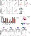
Fig. 1
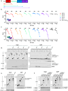
Fig. 1. Proteins required for ZAP antiviral activity.
a Single-cycle HIV-1WT (black) and HIV-1CG (red), infection of HeLa control cells (transduced with empty CRISPR-no guide- vector) multiplicity of infection (MOI) for input virus is plotted on the x-axis and subsequent virus yield from those infected cells (infectious units/mL) is plotted on the y-axis. Data were reported as mean ± standard error of the mean (sem) of three biological replicates. bf Single-cycle infection of ZAP−/− (b) TRIM25−/− (c) KHNYN−/− (d) N4BP1−/− (e) KHNYN−/− N4BP1−/− Hela cells, as in (a). g, h Single-cycle infection of KHNYN−/− N4BP1−/− Hela cells as in (a) following reconstitution with a doxycycline-inducible KHNYN-FLAG (g) or N4BP1-3xHA (h) expression vector. Expression of KHNYN-FLAG or N4BP1-3xHA in the presence or absence of Dox is shown on the right. i EV71WT (dark gray bars) vs EV71CG (red bars) replication in the HeLa cell lines as in (af) above. Luminescence (relative light units) generated by the NanoLuc reporter EV71 viruses measured at 48 h after infection is shown and error bars represent six biological replicates. j Cartoon schematic representation of HEK293T ZAP−/−/TRIM25−/−/KHNYN−/−/N4BP1−/− cell transfection-based reconstitution system to measure virus yield in the presence or absence of ZAP antiviral complex components. kn Reconstitution of ZAP, TRIM25, and KHNYN antiviral activity. Increasing amounts (ng) of plasmids expressing KHNYNWT or KHNYNcat (putative catalytic site mutant) were transfected with 350 ng HIV-1WT or HIV-1CG.proviral plasmids along with empty vector (k), 75 ng TRIM25-expressing plasmid (l), 150 ng ZAP-expressing plasmid (m), or both 150 ng ZAP- and 75 ng TRIM25-expression plasmids (n) Infectious virus yield (in infectious units/mL) from transfected cells measured by infection of MT4 LTR-GFP cells and flow cytometry. Data were reported as mean ± sem of two biological replicates and are representative of three independent experiments.
Fig. 2
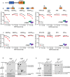
Fig. 2. ZAP domain requirement for CG-specific antiviral activity.
a Schematic depicting ZAP domain organization and truncated proteins used for reconstitution experiments, noting C-terminal domain boundaries. ZAP has an N-terminal RNA-binding domain (RBD, red) containing four zinc fingers (ZnFs), a putatively disordered domain (green), a fifth ZnF, WWE (blue), and PARP (purple) domains are at the C-terminus. b Reconstitution experiments in which increasing amounts of plasmids (ng) expressing the truncated ZAP expression plasmids shown in (a) were co-transfected with a constant amount (75 ng) of KHNYN expression plasmid in 293T ZAP−/− TRIM25−/− KHNYN−/− N4BP1−/− cells. Effects on HIV-1WT and HIV-1CG virus yield was measured as infectious units (IU/ml). Data were reported as mean ± sem of n = 4 biological replicates. c Same as (b) except that 75 ng of a TRIM25 expression plasmid was included in each transfection. d Co-immunoprecipitation of C-terminally 3xHA-tagged ZAP proteins (shown in (a)) transiently transfected in 293 T ZAP−/− TRIM25−/− KHNYN−/− N4BP1−/− cells, with or without doxycycline-induced expression of ZAP-N-FLAG or ZAP-L-FLAG. Proteins were immunoprecipitated from cell lysates with anti-FLAG antibodies and subjected to western blot analysis. ZAP-L with the RNA-binding domain deleted (ZAP-L ΔRBD) was used to evaluate the role of the RBD in ZAP self-association. IP immunoprecipitation, IB immunoblot. e Co-immunoprecipitation of C-terminally 3xHA-tagged ZAP proteins (shown in (a)) transiently transfected in 293T ZAP−/− TRIM25−/− KHNYN−/− N4BP1−/− cells, with doxycycline-induced expression of KHNYN-FLAG. Proteins were immunoprecipitated from cell lysates with anti-FLAG antibodies and subjected to western blot analysis. f Co-immunoprecipitation of TRIM25 with C-terminally 3xHA-tagged ZAP proteins (shown in (a)) transiently transfected in 293 T ZAP−/− TRIM25−/− KHNYN−/− N4BP1−/− cells. Proteins were immunoprecipitated from cell lysates with anti-HA antibody and subjected to western blot analyses with the indicated antibodies.
Fig. 3
Fig. 3. KHNYN domain requirements for complex assembly and antiviral activity.
a Schematic showing KHNYN domain organization with an N-terminal KH domain (blue), a NYN domain (orange), and putative endonuclease catalytic site and a C-terminal domain (CTD. green) with homology to formerly annotated CUBAN domains. b Reconstitution experiments in which increasing amounts of KHNYN expression plasmids (ng) were co-transfected with constant amounts of TRIM25 (75 ng) and ZAP-L (150 ng) expression plasmids. Effects on HIV-1WT and HIV-1CG virus yield was measured as infectious units (IU/ml). Data were reported as mean ± sem of n = 5 biological replicates. c Same as (b) except that the TRIM25 expression plasmid was omitted. d Co-immunoprecipitation of TRIM25 with KHNYN-FLAG following transfection of 293T ZAP−/− TRIM25−/− KHNYN−/− N4BP1−/− cells with plasmids expressing the KHNYN-FLAG proteins described in (a, b). Proteins were immunoprecipitated from cell lysates with an anti-FLAG antibody and subjected to western blot analyses. e Co-immunoprecipitation of ZAP-L-3xHA with KHNYN-FLAG following transfection of 293T ZAP−/− TRIM25−/− KHNYN−/− N4BP1−/− cells with plasmids expressing the KHNYN-FLAG proteins described in (a, b). Proteins were immunoprecipitated from cell lysates with an anti-FLAG antibody and subjected to western blot analyses.
Fig. 4
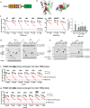
Fig. 4. KHNYN nuclease activity.
a Schematic representation of RNA substrates for KHNYN nuclease assays. Positions of CpG dinucleotides are in red on the CG-5 58-mer (right panel), with corresponding ApA substitutions in white on the CG-0 56-mer (left panel). b, c In vitro nuclease assay using KHNYNWT-FLAG (b) and KHNYNcat-FLAG (D565A/D566A) (c) purified by FLAG affinity chromatography from lysates of 293T ZAP−/− TRIM25−/− KHNYN−/− N4BP1−/− cells Nuclease activity was measured for each protein against the single-stranded CG-0 substrate over a 4-h time course. Activity showed vs a mock reaction with no protein added for comparison.
Fig. 5
Fig. 5. Structure of ZAPX–KHNYNCTD complex.
a Formation of a ZAPX-KHNYNCTD complex. Size-exclusion chromatography profiles of ZAPX (dotted trace), KHNYNCTD (dashed trace) and a 1:2 mixture of ZAPX and KHNYNCTD (solid trace) illustrate the larger complex eluting in the protein mixture, consistent with the SDS-PAGE analysis of fractions from the ZAPX-KHNYNCTD mixture shown below the chromatography profile. Red lines mark the fractions containing the ZAPX-KHNYNCTD complex. One experiment was performed. b Overall structure of the ZAPX-KHNYNCTD complex. ZAPX is shown in blue and KHNYNCTD is shown in purple. Zinc ions are shown as gray spheres. c Details of the interaction between ZAPX (blue) and KHNYNCTD (purple) highlighting the three interacting interfaces. Hydrogen bonds are shown in dashed lines. The zinc (gray sphere) binding site formed by H17 and E29 on ZAPX and H692 on KHNYNCTD is shown with dashed lines. A water molecule bound to zinc is shown as a red sphere. d Reconstitution experiments in which increasing amounts of plasmids (ng) expressing WT and point mutant ZAP proteins informed by the crystal structure were co-transfected with a constant amount (75 ng) of KHNYN expression plasmid in 293T ZAP−/− TRIM25−/− KHNYN−/− N4BP1−/− cells. Effects on HIV-1WT and HIV-1CG virus yield was measured as infectious units (IU/ml). Data were reported as mean ± sem of n = 5 biological replicates, (e) Same as (d) except that 75 ng TRIM25 expression plasmid was also transfected.
Fig. 6
Fig. 6. Molecular determinants of TRIM25 function.
a Schematic representation of TRIM25 highlighting the N-terminal RING domain (orange), two B-box domains (brown), a coiled-coil domain (yellow) that drives inherent dimerization, and a C-terminal SPRY domain (green). b Structure of the TRIM25 RING-domain dimer (from PDB 5EYA) with each subunit displayed in a different gray shade and hydrophobic amino acids at the dimer interface (Leu63, Leu69, and Val72) colored red and green on the respective subunits. c Reconstitution experiments in which increasing quantities of plasmids (ng) expressing wildtype and point mutant TRIM25 were co-transfected with a constant amount of plasmids expressing KHNYN (75 ng) and ZAP-L (150 ng). Wild-type TRIM25 was compared with a deletion of the RING domain, point mutants at hydrophobic residues in the RING domain (L63A, L69A, V72A, and V68A/L69A), a TRIM25 SPRY and proximal 7-Lys (7 K) deletion mutants were tested as well as a 7KA substitution mutant. Effects on HIV-1WT (black) and HIV-1CG (red) virus yield were measured as infectious units (IU/ml). Data were reported as mean ± sem of n = 3 biological replicates. The right panel shows a summary of the relative infectious HIV-1CG virus yield (compared to HIV-1WT) when 100 ng of each variant TRIM25 expression plasmid was transfected. d, e Co-immunoprecipitation of TRIM25 with KHNYN-FLAG (d) or ZAP-L-FLAG (e) following transfection of 293T ZAP−/− TRIM25−/− KHNYN−/− N4BP1−/− cells (stably transduced with doxycycline-inducible KHNYN-FLAG (d) or ZAP-L-FLAG expression constructs (e)) with plasmids expressing TRIM25 domain deletion mutant proteins. Proteins were immunoprecipitated from cell lysates with an anti-FLAG antibody and subjected to western blot analyses. f, g Reconstitution experiments in which increasing amounts of plasmids (ng) expressing TRIM25 whose RING domains (f) or SPRY domains (g) were substituted with the RING domain from the indicated TRIM proteins were co-transfected with a constant amount of plasmids expressing KHNYN (75 ng) and ZAP-L (150 ng). Effects on HIV-1WT (black) and HIV-1CG (red) virus yield was measured as infectious units (IU/ml). Data were reported as mean ± sem of n = 4 biological replicates.
Fig. 7
Fig. 7. Reconstitution of antiviral activity with designed chimeric antiviral proteins.
a Schematic representation of a ZAP-KHNYN chimera constructed by fusing the ZAP RNA binding domain (RBD) to the catalytic NYN domain from KHNYN. b Reconstitution experiments in which 293T ZAP−/− TRIM25−/− KHNYN−/− N4BP1−/− cells were transfected with increasing amounts of plasmids (ng) expressing the ZAPRBD-NYN chimera in the absence of KHNYN or ZAP, in the presence or absence of a TRIM25 expression plasmid (75 ng). Effects on HIV-1WT (black) and HIV-1CG (red) virus yield was measured as infectious units (IU/ml). Data were reported as mean ± sem of n = 4 biological replicates. c Lysates from 293T ZAP−/− TRIM25−/− KHNYN−/− N4BP1−/− cells, transfected as in Fig. 7b were analyzed by western blotting probing with antibodies against RBD-NYN-FLAG, HIV-1 gp120, and GAPDH. d Schematic representation of a TRIM25/ZAP chimera constructed by fusing the N-terminus of TRIM25 (RING-B-box-Coiled-Coil) with the ZAP RBD. e Reconstitution experiments in which 293T ZAP−/− TRIM25−/− KHNYN−/− N4BP1−/− cells were transfected with increasing amounts of plasmids (ng) expressing the TRIM25-ZAPRBD chimera plus KHNYN in the absence of ZAP or TRIM25. For comparison, cells were transfected with ZAP-N and ZAP-L, plus TRIM25 and KHNYN, or KHNYN alone. Effects on HIV-1WT (black) and HIV-1CG (red) virus yield was measured as infectious units (IU/ml). Data were reported as mean ± sem of n = 4 biological replicates. f Lysates from 293T ZAP−/− TRIM25−/− KHNYN−/− N4BP1−/− cells, transfected as in Fig. 7e were analyzed by western blotting probing with antibodies against TRIM25-RBD-3xHA and Tubulin.
Fig. 8
Fig. 8. Model for mechanism of action of ZAP, KHNYN, and TRIM25 in CpG-specific antiviral activity.
a Model for the assembly of ZAP, KHNYN (or N4BP1), TRIM25 and a target RNA that enables the nuclease to specifically deplete CpG RNAs that are recognized through multivalent interaction with ZAP-containing complexes whose multimerization is driven by ZAP:ZAP, ZAP:TRIM25, and TRIM25:TRIM25 interactions. b Details of the interactions between the core functional ZAP NTD a CpG dinucleotide containing RNA and the KHNYN CTD that enable monovalent recruitment of a KHNYN nuclease to a target RNA, (c) Details of the TRIM25 RING-RING interaction that contribute to the formation of ZAP containing complexes that recognize CpG-rich RNA in a multivalent manner.

References

    1. Lai, W. S., Wells, M. L., Perera, L. & Blackshear, P. J. The tandem zinc finger RNA binding domain of members of the tristetraprolin protein family. Wiley Interdiscip. Rev. RNA10, e1531 (2019). - PMC - PubMed
    1. Uehata, T. & Akira, S. mRNA degradation by the endoribonuclease Regnase-1/ZC3H12a/MCPIP-1. Biochim. Biophys. Acta1829, 708–713 (2013). - PubMed
    1. Gao, G., Guo, X. & Goff, S. P. Inhibition of retroviral RNA production by ZAP, a CCCH-type zinc finger protein. Science297, 1703–1706 (2002). - PubMed
    1. Guo, X., Carroll, J. W., Macdonald, M. R., Goff, S. P. & Gao, G. The zinc finger antiviral protein directly binds to specific viral mRNAs through the CCCH zinc finger motifs. J. Virol.78, 12781–12787 (2004). - PMC - PubMed
    1. Bick, M. J. et al. Expression of the zinc-finger antiviral protein inhibits alphavirus replication. J. Virol.77, 11555–11562 (2003). - PMC - PubMed

Publication types

MeSH terms

Associated data

LinkOut - more resources