. 2024 Dec 30;15(1):10804.
doi: 10.1038/s41467-024-55072-6.

Artificial intelligence-driven rational design of ionizable lipids for mRNA delivery

Affiliations

Artificial intelligence-driven rational design of ionizable lipids for mRNA delivery

Wei Wang et al. Nat Commun. .

Abstract

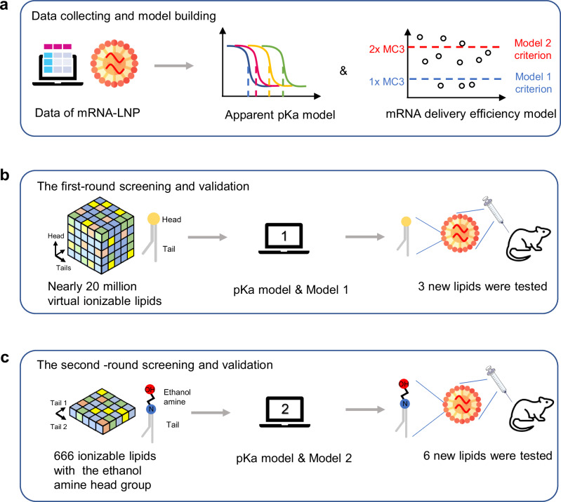
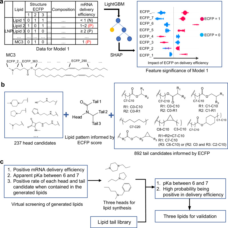
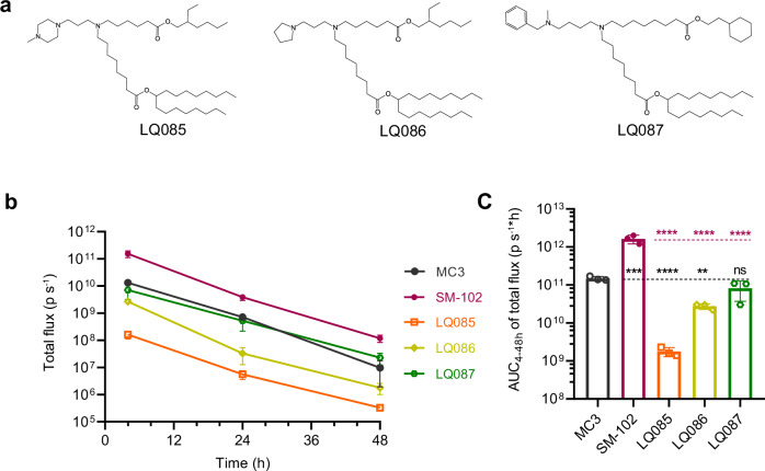
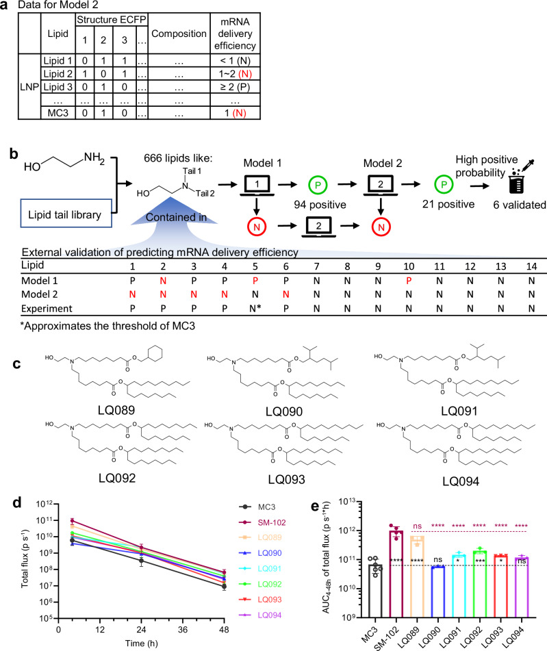
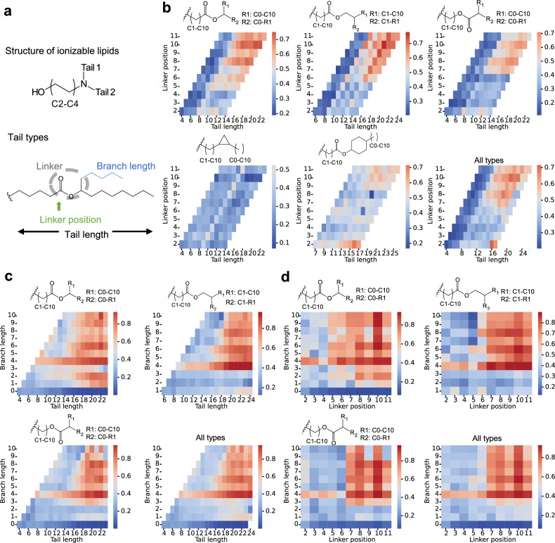
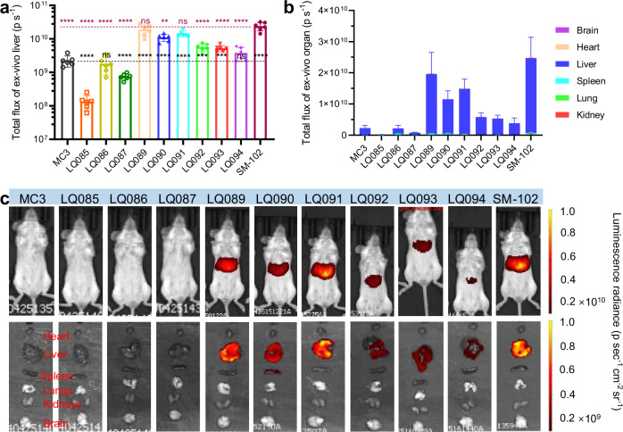
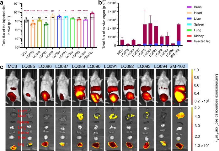
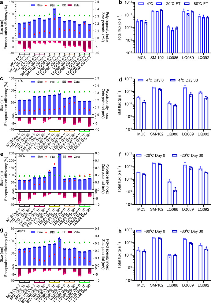
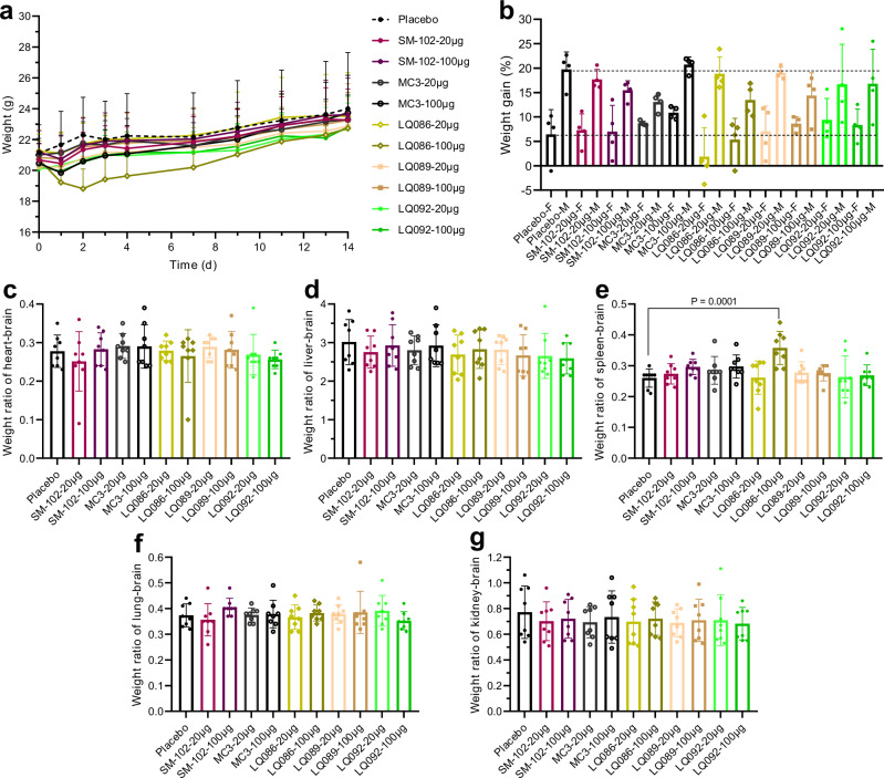
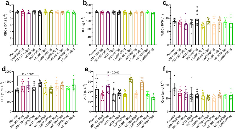
Lipid nanoparticles (LNPs) have proven effective in mRNA delivery, as evidenced by COVID-19 vaccines. Its key ingredient, ionizable lipids, is traditionally optimized by inefficient and costly experimental screening. This study leverages artificial intelligence (AI) and virtual screening to facilitate the rational design of ionizable lipids by predicting two key properties of LNPs, apparent pKa and mRNA delivery efficiency. Nearly 20 million ionizable lipids were evaluated through two iterations of AI-driven generation and screening, yielding three and six new molecules, respectively. In mouse test validation, one lipid from the initial iteration, featuring a benzene ring, demonstrated performance comparable to the control DLin-MC3-DMA (MC3). Notably, all six lipids from the second iteration equaled or outperformed MC3, with one exhibiting efficacy akin to a superior control lipid SM-102. Furthermore, the AI model is interpretable in structure-activity relationships.


Conflict of interest statement

Competing interests: The authors declare no competing interests.

Figures

Fig. 1
Fig. 1. Overview of AI-driven rational design of ionizable lipids for mRNA lipid nanoparticles.
a The collected data was used to build models predicting the apparent pKa and mRNA delivery efficiency of LNPs. The 1- and 2-fold of MC3 mRNA delivery efficiency was used in Models 1 and 2 as the criterion in the model classifying ionizable lipids with positive delivery efficiency. b The first-round of virtual screening of ionizable lipids and validation based on pKa model and Model 1. c The second-round of virtual screening of ionizable lipids and validation based on pKa model and Model 2. AI artificial intelligence, LNP lipid nanoparticle, MC3, DLin-MC3-DMA.
Fig. 2
Fig. 2. Overview of the first-round lipid virtual screening.
a Data representation of the AI model (Model 1), and the methods of model training (LightGBM) and feature significance calculation (SHAP). Some typical ECFP bits and their corresponding substructure of MC3 are shown as an example. P, positive; N, negative. b Significant substructures informed by the model and used for lipid generation. c The method of lipid virtual screening and three lipids were selected for experimental validation. ECFP extended connectivity fingerprints, LightGBM Light Gradient Boosting Machine, SHAP SHapley Additive exPlanations, MC3 DLin-MC3-DMA, AI artificial intelligence.
Fig. 3
Fig. 3. Experimental validation of the three ionizable lipids resulted from the first-round of virtual screening.
a The structure of the three ionizable lipids selected from the first round of virtual screening. b Female BALB/c mice (6-8 weeks old) were intravenously injected with LNPs loaded with luciferase mRNA at a dose of 5 μg per mouse, and at certain time points, total luminescence was detected after injection of D-luciferin. c The AUC of total luminescence. All data are presented as the mean ± SD (n = 3). Statistical significance was analyzed by one-way ANOVA (ns, not significant; *p < 0.0332; **p < 0.0021; ***p < 0.0002; ****p < 0.0001. The P makers in black are results of the comparisons were with MC3, and those in red are with SM-102. Source data are provided as a Source Data file.). MC3 DLin-MC3-DMA, AUC area under curve, LNP lipid nanoparticle, SD standard deviation, ANOVA Analysis of Variance.
Fig. 4
Fig. 4. Overview of the second-round lipid virtual screening and experimental validation.
a Data representation of Model 2 predicting mRNA delivery efficiency. Compared to the representation method of Model 1, the positive criterion was set as 2-fold the delivery efficiency of the standard MC3 LNP. b External validation of Model 2, and associating Model 1 and 2 to screen the generated ionizable lipids. c The six lipids selected for experimental validation. d Female BALB/c mice (6–8 weeks old) were intravenously injected with LNPs loaded with luciferase mRNA at a dose of 5 μg per mouse, and at certain time points, total luminescence was detected after injection of D-luciferin. Time courses of the total flux of the screened six lipids. e The AUC of total luminescence of the screened six lipids. For MC3, SM-102, and other groups, n = 6, 5, 3, respectively. All data are presented as the mean ± SD. Statistical significance was analyzed by one-way ANOVA (ns, not significant; *p < 0.0332; **p < 0.0021; ***p < 0.0002; ****p < 0.0001. The P makers in black are results of the comparisons with MC3, and those in red are with SM-102. Source data are provided as a Source Data file). MC3 DLin-MC3-DMA, AUC area under curve, LNP lipid nanoparticle, SD standard deviation, ANOVA Analysis of Variance.
Fig. 5
Fig. 5. Structure analysis of ionizable lipids informed by AI models.
a Ionizable lipid pattern and tail types for analysis. bd Heatmap of positive rates for specific tail types. All types of tails and heads containing hydroxyl were combined to form ionizable lipids in an exhaustive manner. Their mRNA delivery efficiency and apparent pKa were predicted with Model 1. For each type of tail, the number of resulting lipids containing the tail and among them the number of positive lipids (efficiency higher than the standard MC3 formulation and pKa between 6.0 and 7.0) were used to calculate the positive rate. The structure-activity relationship is shown as the influence of tail length and linker position (b) tail length and branch length (c), and the branch length and linker position (d) on the positive rate. AI, artificial intelligence.
Fig. 6
Fig. 6. Luminescence distribution and expression of luciferase mRNA loaded in different LNPs via intravenous administration.
a Liver luminescence of luciferase at 4 h. b Organ-distributed luminescence of luciferase at 4 h. (c) Representative images of the luminescence. Each group had three female BALB/c mice and three male ones. All data are presented as the mean ± SD. Statistical significance was analyzed by one-way ANOVA (ns, not significant; *p < 0.0332; **p < 0.0021; ***p < 0.0002; ****p < 0.0001. The P makers in black are the results of the comparisons with MC3, and those in red are with SM-102. Source data are provided as a Source Data file.). MC3 DLin-MC3-DMA, SD standard deviation, ANOVA Analysis of Variance.
Fig. 7
Fig. 7. Luminescence distribution and expression of luciferase mRNA loaded in different LNPs via intramuscular administration.
a Luminescence of the injection site at 4 h. b Organ-distributed luminescence of luciferase at 4 h. c Representative images of the luminescence. Each group had three female BALB/c mice. All data are presented as the mean ± SD. Statistical significance was analyzed by one-way ANOVA (ns, not significant; *p < 0.0332; **p < 0.0021; ***p < 0.0002; ****p < 0.0001. The P makers in black are results of the comparisons were with MC3, and those in red are with SM-102. Source data are provided as a Source Data file.). MC3 DLin-MC3-DMA, SD standard deviation, ANOVA Analysis of Variance.
Fig. 8
Fig. 8. LNP stability during the freezing-thawing process and long-term storage.
a, b Changes in particle characteristics and in vivo efficiency before and after the freezing-thawing process. c, d Changes in particle characteristics and in vivo efficiency during long-term storage at 4 °C. e, f Changes in particle characteristics and in vivo efficiency during long-term storage at −20 °C. g, h Changes in particle characteristics and in vivo efficiency during long-term storage at −80 °C. The small colored lines beneath the X-axes in panels a, c, e and g are used to distinguish the lipid groups more clearly. For in vivo efficiency of each group, three female BALB/c mice were intravenously administered luciferase mRNA loaded in LNPs at a dosage of 5 μg per mouse and the luminescence signal of the whole body was detected 4 hours later post intraperitoneal administration of D-luciferin. All data are presented as the mean ± SD (n = 3). The analysis of change trends met the needs of the stability study, so significance analysis was not performed. Source data are provided as a Source Data file. MC3 DLin-MC3-DMA, PDI polydispersity index, EE encapsulation efficiency, LNP lipid nanoparticle, SD standard deviation.
Fig. 9
Fig. 9. Weight monitoring and organ coefficient in the acute toxicity test.
a Weight-time curve. The placebo was saline. Each group had four female BALB/c mice and four male ones. b Weight gain at Day 14 compared to Day 0. F=female, M=male. cg Coefficient of heart-brain, liver-brain, spleen-brain, lung-brain and kidney-brain, respectively. All data are presented as the mean ± SD (n = 8 for each dose, including four females and four males). Statistical significance was analyzed by one-way ANOVA (unmarked, not significant. Source data are provided as a Source Data file.). MC3 DLin-MC3-DMA, SD standard deviation, ANOVA Analysis of Variance.
Fig. 10
Fig. 10. Hematological indices.
af Blood was obtained 14 days after intravenous administration. All data are presented as the mean ± SD (n = 8 for each dose, including four females and four males). Statistical significance was analyzed by one-way ANOVA. (Unmarked, not significant. Source data are provided as a Source Data file.). MC3 DLin-MC3-DMA, RBC red blood cell, HGB hemoglobin, WBC white blood cell, PLT platelet, ALT glutamic-pyruvic transaminase, Crea creatinine, SD standard deviation, ANOVA Analysis of Variance.

Similar articles

Cited by

References

    1. Baden Lindsey, R. et al. Efficacy and Safety of the mRNA-1273 SARS-CoV-2 Vaccine. N. Engl. J. Med.384, 403–416 (2021). - PMC - PubMed
    1. Polack, F. P. et al. Safety and Efficacy of the BNT162b2 mRNA Covid-19 Vaccine. N. Engl. J. Med.383, 2603–2615 (2020). - PMC - PubMed
    1. Hou, X., Zaks, T., Langer, R. & Dong, Y. Lipid nanoparticles for mRNA delivery. Nat. Rev. Mater.6, 1078–1094 (2021). - PMC - PubMed
    1. World Health Organization. COVID-19 vaccine tracker and landscape. https://www.who.int/publications/m/item/draft-landscape-of-covid-19-cand... (2022).
    1. Semple, S. C. et al. Efficient encapsulation of antisense oligonucleotides in lipid vesicles using ionizable aminolipids: formation of novel small multilamellar vesicle structures. Biochim. Biophys. Acta BBA - Biomembr.1510, 152–166 (2001). - PubMed

Publication types

LinkOut - more resources