. 2024 Dec 30;15(1):10741.
doi: 10.1038/s41467-024-54960-1.

Minimal presynaptic protein machinery governing diverse kinetics of calcium-evoked neurotransmitter release

Affiliations

Minimal presynaptic protein machinery governing diverse kinetics of calcium-evoked neurotransmitter release

Dipayan Bose et al. Nat Commun. .

Abstract

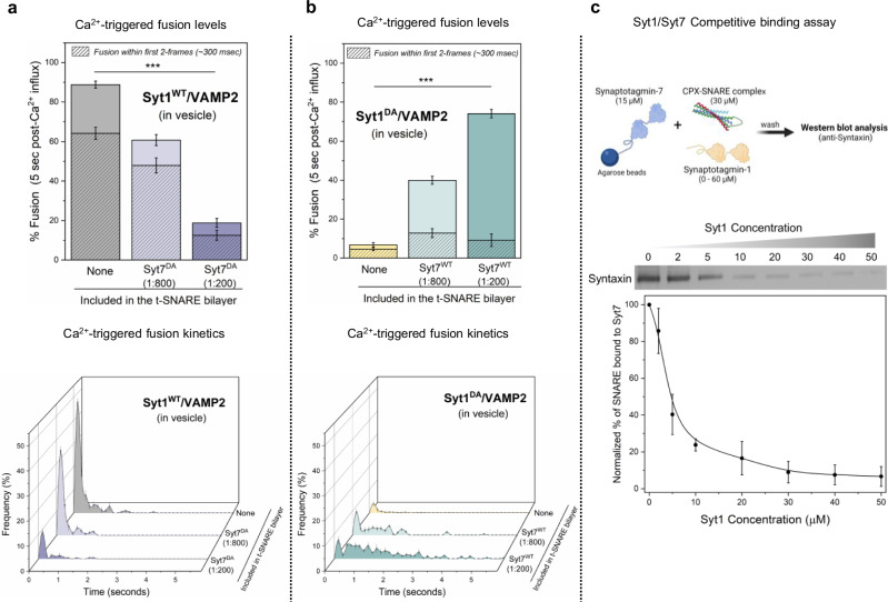
Neurotransmitters are released from synaptic vesicles with remarkable precision in response to presynaptic calcium influx but exhibit significant heterogeneity in exocytosis timing and efficacy based on the recent history of activity. This heterogeneity is critical for information transfer in the brain, yet its molecular basis remains poorly understood. Here, we employ a biochemically-defined fusion assay under physiologically relevant conditions to delineate the minimal protein machinery sufficient to account for various modes of calcium-triggered vesicle fusion dynamics. We find that Synaptotagmin-1, Synaptotagmin-7, and Complexin synergistically restrain SNARE complex assembly, thus preserving vesicles in a stably docked state at rest. Upon calcium activation, Synaptotagmin-1 induces rapid vesicle fusion, while Synaptotagmin-7 mediates delayed fusion. Competitive binding of Synaptotagmin-1 and Synaptotagmin-7 to the same SNAREs, coupled with differential rates of calcium-triggered fusion clamp reversal, govern the overall kinetics of vesicular fusion. Under conditions mimicking sustained neuronal activity, the Synaptotagmin-7 fusion clamp is destabilized by the elevated basal calcium concentration, thereby enhancing the synchronous component of fusion. These findings provide a direct demonstration that a small set of proteins is sufficient to account for how nerve terminals adapt and regulate the calcium-evoked neurotransmitter exocytosis process to support their specialized functions in the nervous system.


Conflict of interest statement

Competing interests: The authors declare no competing interests.

Figures

Fig. 1
Fig. 1. Impact of Syt7 on Ca2+-evoked fusion of Syt1/VAMP2 vesicles.
a In a typical in vitro fusion experiment, vesicles containing VAMP2 (~70 copies) and Syt1 (~20 copies) were added to a suspended bilayer membrane (formed on a silicon substrate with 5 μm holes) reconstituted with Syntaxin/SNAP25 (1:400 protein-to-lipid ratio) ±Syt7 in the presence of Complexin (2 µM) in solution. The fate of each vesicle before and after the addition of 100 µM Ca2+ was monitored by a confocal microscope using a fluorescent (ATTO647N) marker included in the vesicle. b Syt7 (included in the t-SNARE containing bilayer) had no impact on the fusion competence of docked Syt1/VAMP2 vesicles, with ~85% fusing within 5 s after the arrival of 100 μM Ca2+ signal at or near the docked vesicles. The hatched bar represents the percent fusion occurring with 2 frames (~300 ms) following Ca2+ arrival. c Syt7 altered the Ca2+-triggered fusion kinetics of docked Syt1/VAMP2 vesicles. Top, Representative time-lapse image of Ca2+-evoked fusion of docked vesicles shows that without Syt7 the vesicles fuse rapidly and synchronously following Ca2+ addition. The inclusion of Syt7 (1:200 protein-to-lipid ratio) introduces variable delays in Ca2+-evoked fusion kinetics. Individual vesicles (white circles) docked within 5 µm suspended bilayer are shown. Bottom, quantitative analysis of Ca2+-evoked fusion of Syt1/VAMP2 vesicles shows that Syt7 introduces a concentration-dependent delay in the Ca2+-evoked fusion kinetics, resulting in a significant reduction in the proportion of vesicles fusing within the first 2 frames (~300 ms) following Ca2+ arrival at time t = 0 s. Data (mean ± standard deviation) are from 5 independent experiments (N = 5) for each condition (~40–50 vesicles per experiment). One-way ANOVA revealed statistically significant difference (***p < 0.001) in Ca2+-coupled fusion occurring within ~300 ms (hatched bar) between groups. The data from ANOVA and Tukey’s HSD post-hoc comparing specific groups is shown in Supplementary Table 1. The source data is provided as a ‘Source Data’ file. Figure 1a created in BioRender. Krishnakumar, S. (2023) BioRender.com/f92r389.
Fig. 2
Fig. 2. Contribution of Syt7 to the establishment of the fusion clamp.
The involvement of Syt7 in the fusion clamp was evaluated using vesicles containing low-copy VAMP2 (~15 copies) and a non-clamping Syt1 mutant, Syt1Q (carrying R281A,E295A,Y338W,R398A,R399A mutations that disrupt the Syt1-SNARE primary interface) in the absence of CPX. a The time between docking and spontaneous fusion was measured for each docked vesicle and the ‘docking-to-fusion’ latency time was cumulatively expressed as the survival percentage. This ‘survival analysis’ provided the measure of the strength of the fusion clamp. In the absence of Syt7 (gray), the majority of the docked VAMP2low/Syt1Q vesicles proceed to fuse spontaneously with a half-time of ~1 s. The inclusion of Syt7 in the bilayer resulted in stably docked vesicles in an immobile state, with clamping efficiency correlating with the amount of Syt7 included. Approximately 40% of vesicles were clamped under low Syt7 concentration (1:800, light blue) and this increased to ~90% under high Syt7 concentration (1:200, dark blue). b, c Syt7 clamped VAMP2low/Syt1Q vesicles remained fusion competent and could be triggered to fuse by the addition of Ca2+ (100 µM) and the observed fusion was desynchronized to the Ca2+ signal. In the absence of Syt7, a very small percent of the docked VAMP2low/Syt1Q vesicles underwent fusion which precluded meaningful kinetic analysis. Data (mean ± standard deviation) are from 3 independent experiments (N = 3) for each condition (~40–50 vesicles per experiment). One-way ANOVA revealed statistically significant difference (***p < 0.001) in % Ca2+-evoked fusion of docked vesicles in the presence of Syt7 as compared to the condition without Syt7 in the bilayer. The data from ANOVA and Tukey’s HSD post-hoc comparing specific groups are shown in Supplementary Table 2. The source data is provided as a ‘Source Data’ file.
Fig. 3
Fig. 3. Syt1 and Syt7 synergistically regulate Ca2+-evoked fusion via competitive binding to the same SNARE complex.
a The inclusion of the Ca2+ binding deficient Syt7 mutant (Syt7DA) in the bilayer inhibited Ca2+ (100 µM) evoked fusion of Syt1WT/VAMP2 vesicles in a dose-dependent manner, without altering the overall fusion kinetics. b Syt7WT from the bilayer rescued the Ca2+-evoked fusion of Syt1DA /VAMP2 vesicles but the fusion events were desynchronized to the Ca2+ signal. Complexin (2 µM) in solution was included in all experiments. c Quantitative pull-down and Western-blot analysis with Syt7 as ‘bait’ and CPX-SNARE complex as ‘prey’ demonstrate that Syt1 disrupts Syt7-SNARE interaction in a concentration-dependent manner. Data (mean ± standard deviation) are from 5 independent experiments (N = 5) for each condition (~40–50 vesicles per experiment) in (a) and (b) and from 4 independent experiments (N = 4) in (c). One-way ANOVA revealed statistically significant difference in % Ca2+-evoked fusion of docked vesicles in the presence of Syt7DA (***p < 0.001) or Syt7WT (***p < 0.001) as compared to condition without Syt7 in the bilayer. The data from ANOVA and Tukey’s HSD post-hoc comparing specific groups are shown in Supplementary Tables 3 and 4 respectively. The source data is provided as a ‘Source Data’ file. Figure 3c (top) created in BioRender. Krishnakumar, S. (2023) BioRender.com/p26b995.
Fig. 4
Fig. 4. Computational model of synergistic activation of vesicular fusion by Syt1 and Syt7.
a Schematic illustration of the release of inhibition model. At rest, fusion of vesicles is inhibited (‘clamped’) by binding of Syt1 and Syt7 along with CPX to partially assembled SNAREpins. Upon Ca2+ binding, the C2 domains of Syt1 and Syt7 insert into the membrane, leading to the removal of the fusion clamp. This allows the complete zippering of the SNARE complexes, resulting in vesicular fusion. Inset shows that two clamp architectures are considered in the default model: dual Syt1/Syt1 or dual Syt1/Syt7 clamp (see Supplementary Fig. 6 for additional clamp architectures tested). b Kinetic reaction schemes describing Ca2+-triggered release of the fusion clamp. Each modeled C2 domain sequentially binds two Ca2+ ions which triggers the insertion of its aliphatic loop into the membrane. Scheme 1 assumes that membrane insertion results in the instantaneous removal of the Synaptotagmin fusion clamp, while Scheme 2 assumes a delay between membrane insertion and the removal of the clamp. S0, S1, S2 refer to 0, 1 or 2 Ca2+ bound state of the C2 domains, while I2 refers to membrane inserted state of the Ca2+-bound C2 domain. The prefixes c and u refer to the ‘clamped’ or ‘unclamped’ state respectively. c The time course of vesicular fusion (Model Output) simulated in response to the experimentally constrained Ca2+ signal (Supplementary Fig. 3) for models with different clamp architecture and kinetics of clamp reversal. Experimental data (mean ± standard deviation from Fig. 1c) for the Ca2+-triggered fusion of Syt1 containing vesicles in the absence (Syt1EXP) or the presence of saturating levels of Syt7 (Syt7EXP) are plotted for comparison. The model suggests that experimentally observed fusion kinetics can be explained by the mechanism with differential rates of fusion clamp removal for Syt1 (instantaneous) and Syt7 (delayed). For each modeled condition a minimum of 1000 stochastic simulations were performed to calculate the average response. The source data is provided as a ‘Source Data’ file. Figure 4a created in BioRender. Krishnakumar, S. (2023) BioRender.com/x49d271.
Fig. 5
Fig. 5. Syt7 enhances Ca2+-synchronized fusion under elevated [Ca2+]basal conditions.
a Comparison of the Ca2+ (100 µM) evoked fusion characteristics without (green) or with (pink) 0.5 μM [Ca2+]basal included during vesicle docking reveals that when pre-activated, Syt7 (included in the bilayer at 1:200 protein-to-lipid ratio) increases the proportion of Ca2+-coupled release of Syt1-containing vesicles (Syt1WT/Syt7WT) without changing the overall fusion levels. This enhancement was not observed with Syt1WT alone. Notably, a similar degree of enhancement of Ca2+-synchronized release was observed with vesicles containing Ca2+-binding deficient Syt1DA (Syt1DA/Syt7WT). Data (mean ± standard deviation) are from 5 independent experiments (N = 5) for each condition (~40–50 vesicles per experiment). Complexin (2 µM) in solution was included in all experiments. b The dual Syt1/Syt7 clamp model, incorporating the delayed release of the clamp for Syt7, reproduces the experimentally observed enhancement of synchronous release following pre-activation with low micromolar [Ca2+]. The extent of facilitation correlated with the level of pre-activating [Ca2+] used. For each modeled condition a minimum of 1000 stochastic simulations were performed to compute the average response. ***p < 0.001 using the one-sided students’ t-test comparison to the control condition with no [Ca2+]basal. The source data is provided as a ‘Source Data’ file.

Similar articles

References

    1. Kaeser, P. S. & Regehr, W. G. Molecular mechanisms for synchronous, asynchronous, and spontaneous neurotransmitter release. Annu Rev. Physiol.76, 333–363 (2014). - PMC - PubMed
    1. Sudhof, T. C. Neurotransmitter release: the last millisecond in the life of a synaptic vesicle. Neuron80, 675–690 (2013). - PMC - PubMed
    1. Regehr, W. G. Short-term presynaptic plasticity. Cold Spring Harb. Perspect. Biol.4, a005702 (2012). - PMC - PubMed
    1. Jackman, S. L. & Regehr, W. G. The Mechanisms and Functions of Synaptic Facilitation. Neuron94, 447–464 (2017). - PMC - PubMed
    1. Reyes, A. et al. Target-cell-specific facilitation and depression in neocortical circuits. Nat. Neurosci.1, 279–285 (1998). - PubMed

Publication types

MeSH terms

LinkOut - more resources