. 2024 Dec 30;15(1):10906.
doi: 10.1038/s41467-024-55268-w.

Mechanisms of recalcitrant fucoidan breakdown in marine Planctomycetota

Affiliations

Mechanisms of recalcitrant fucoidan breakdown in marine Planctomycetota

Carla Pérez-Cruz et al. Nat Commun. .

Abstract

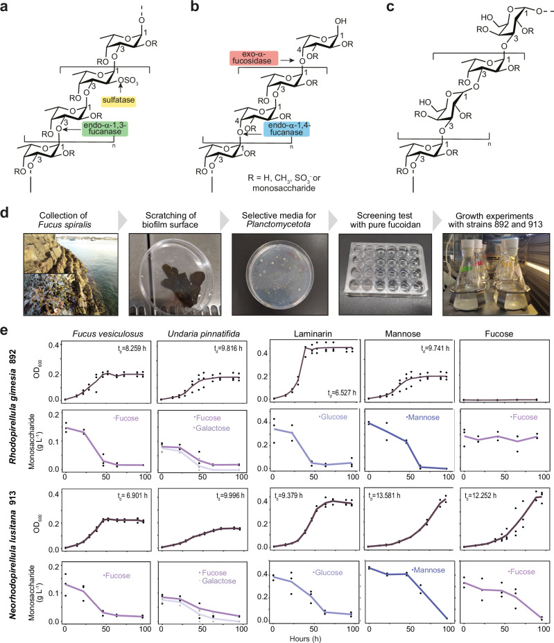
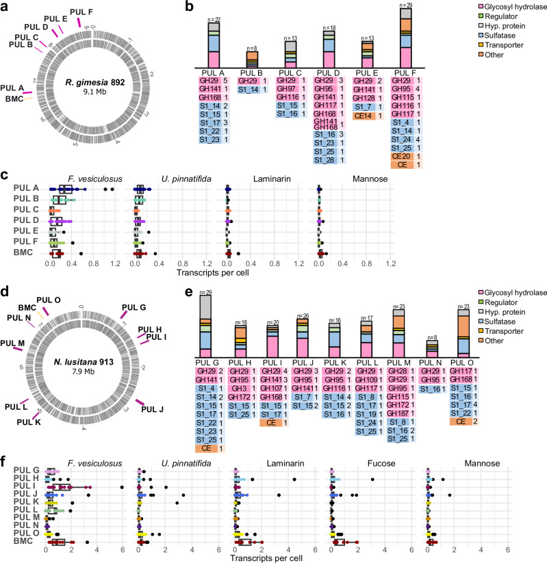
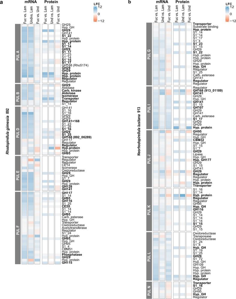
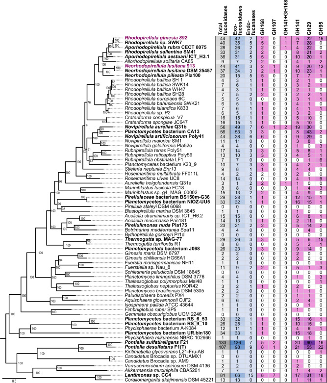
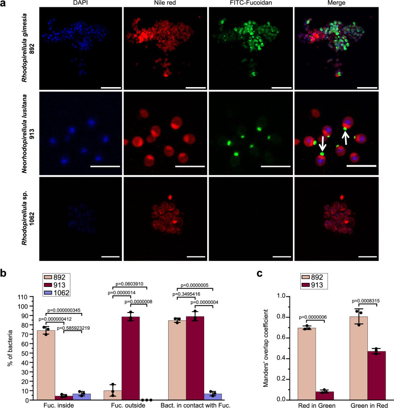
Marine brown algae produce the highly recalcitrant polysaccharide fucoidan, contributing to long-term oceanic carbon storage and climate regulation. Fucoidan is degraded by specialized heterotrophic bacteria, which promote ecosystem function and global carbon turnover using largely uncharacterized mechanisms. Here, we isolate and study two Planctomycetota strains from the microbiome associated with the alga Fucus spiralis, which grow efficiently on chemically diverse fucoidans. One of the strains appears to internalize the polymer, while the other strain degrades it extracellularly. Multi-omic approaches show that fucoidan breakdown is mediated by the expression of divergent polysaccharide utilization loci, and endo-fucanases of family GH168 are strongly upregulated during fucoidan digestion. Enzymatic assays and structural biology studies reveal how GH168 endo-fucanases degrade various fucoidan cores from brown algae, assisted by auxiliary hydrolytic enzymes. Overall, our results provide insights into fucoidan processing mechanisms in macroalgal-associated bacteria.


Conflict of interest statement

Competing interests: The authors declare no competing interests.

Figures

Fig. 1
Fig. 1. Chemical structure of fucoidans and experimental work with Planctomycetota strains isolated from Fucus spiralis.
Chemical structures of the common backbone chains of Type I (a) and Type II (b) homofucans, including the target site for the fucoidan-processing enzymes. c Chemical structure of the common backbone of the galactofucan (heterofucan) found in Undaria pinnatifida. d Workflow of the experimental work. e Growth curves and carbohydrate consumption of strains 892 and 913 in different carbon sources (i.e., fucoidan from Fucus vesiculosus and Undaria pinnatifida, laminarin, mannose and fucose). Data points at each time represent three biologically independent experiments. Doubling times (td) have been derived from optical density measurement values (OD600) during exponential phase. Source data are provided as a Source Data file.
Fig. 2
Fig. 2. Fucoidan Polysaccharide Utilization Loci (PULs) in Planctomycetota strains and global expression under different carbon sources obtained by quantitative transcriptomics.
Genomic location of putative fucoidan PULs in strains 892 (a) and 913 (d) and functional composition of the PULs (b and e). Besides endo-fucanases (GH107, GH168 and GH187) and exo-fucosidases (GH29, GH95, GH141), all indicated in pink color, we identified enzymes from families GH97 (α-glucosidase/α-galactosidase), GH115 (α-glucuronidase), GH116 (β-glucosidase), GH117 (α-neoagarobiosidase) and GH128 (endo-β−1,3-glucanase/ exo-β−1,3-glucanase) in the PULs of strain 892 (b), and enzymes from families GH3 (β-glucosidase), GH28 (endo-polygalacturonase/exo-polygalacturonase), GH109 (α-N-acetylgalactosaminidase), GH115 (α-glucuronidase), GH116 (β-glucosidase), GH117 (α-neoagarobiosidase), GH172 (α-d-arabinofuranosidase) in the PULs of strain 913 (e). Pannels c and f show mean transcripts per cell of individual genes in each of the fucoidan PULs and the BMC (highlighted in different colors) in strains 892 and 913, respectively, grown on different carbon sources (i.e., fucoidan from Fucus vesiculosus and Undaria pinnatifida, laminarin, mannose and fucose) in three biologically independent experiments. Box plots indicate median (middle line), 25th, 75th percentile (box), and 5th and 95th percentile (whiskers). Individual plotted data points represent the transcripts per cell and black points indicate the outliers. Transcripts per cell were quantified by transcriptomics using internal mRNA standards in three biologically independent replicates. Source data are provided as a Source Data file. BMC: Bacterial microcompartment; GH: glycosyl hydrolase; S: sulfatase; CE: carbohydrate esterase.
Fig. 3
Fig. 3. Differential gene expression at mRNA and protein level in the identified fucoidan PULS from strains 892 and 913.
Log2-fold changes (LFC) of mRNA and protein expression data are based on pairwise comparisons between different carbon sources in strains 892 (a) and 913 (b). Color scale indicates upregulation (blue) or downregulation (red) of transcripts or proteins in the indicated fucoidan vs. other carbon sources. Expression values were quantified in three biologically independent replicates (n = 3). Only PULs where fucosidases were detected at the protein level are shown. Bold letters highlight those proteins that were significantly more abundant in one of the fucoidan sources as compared to laminarin (two-sided Student’s t-test, p value < 0.05). Source data are provided as a Source Data file. Fuc: fucoidan from Fucus vesiculosus; Und: fucoidan from Undaria pinnatifida; Lam: laminarin; GH: glycosyl hydrolase; S: sulfatase; CE: carbohydrate esterase; CBM: carbohydrate binding modules; Hyp: hypothetical; Carb: carbohydrate.
Fig. 4
Fig. 4. Endo-fucanases and exo-fucosidases detected in selected genomes from Planctomycetota and Verrucomicrobiota phyla.
The recently characterized endo-fucanase families GH187 and GH174 were not included in this comparative analysis since the assignation was not found to be reliable using dbCAN v3.0.7. Taxa enriched in fucosidases, i.e., displaying ≥ 20 fucosidases, are highlighted in bold. Strains 892 and 913 are highlighted in magenta. The phylogenetic tree on the left side shows the taxonomic profiling of the selected genomes, based on the amino acid sequences of 10 core genes aligned using the Phylogenetic Tree Service from BV-BRC database (https://www.bv-brc.org) without deletion and duplication allowed, using Candidatus Brocadiia as outgroup. Enzymes with more than one domain have not been considered in the total sum of fucosidases. Source data are provided as a Source Data file.
Fig. 5
Fig. 5. Phylogenetic tree of GH168 homologs.
Maximum likelihood phylogenetic tree of GH168 homolog amino acid sequences from all available Planctomycetota genomes at NCBI, selected genomes of Bacteroidota and Verrucomicrobiota, and environmental sequences found in the The Ocean Microbiomics Database gene catalog. Source data are provided as a Source Data file. The number of collapsed nodes are indicated in parentheses. The GH168 enzymes of strains 892 and 913 are highlighted in magenta. The GH168 enzymes Rho5174 and PbFucA, which are the focus of this study, are highlighted with star symbols.
Fig. 6
Fig. 6. Hydrolytic activity of PbFucA, Rho5174 and strain 892 lysates against fucoidan from F. vesiculosus and U. pinnatifida.
Hydrolytic activity of Rho5174 (left) or PbFucA (right) against fucoidan from F. vesiculosus (a) and U. pinnatifida (b). The hydrolytic activity of the enzymes, strain 892 lysate grown in F. vesiculosus (L892-Fv, in panel a) or U. pinnatifida (L892-Up, in panel b), and the combination of enzyme and lysate are colored in gray, cyan, and magenta, respectively. Data represent the mean ± SD derived from three biologically independent experiments (n = 3). c C-PAGE analysis of the hydrolytic activity of Rho5174 and PbFucA, strain 892 lysate grown in F. vesiculosus (L892-Fv), and the combination of enzymes and lysate against fucoidan from F. vesiculosus. d C-PAGE analysis of the hydrolytic activity of Rho5174 and PbFucA, strain 892 lysate grown in U. pinnatifida (L892-Up) and the combination of enzymes and lysate against fucoidan from U. pinnatifida. Source data are provided as a Source Data file.
Fig. 7
Fig. 7. The overall structure of Rho5174.
a Cartoon representation showing the general fold and secondary structure organization of Rho5174. Domains, MOPS molecule (MPO) and catalytic residues are annotated. b Surface representation of the Rho5174. Loops that form the main groove of Rho5174 are highlighted in different colors. c Electrostatic surface representations of Rho5174 showing the location of the catalytic site. d–f Close-up view of the catalytic active site of Rho5174 shown as cartoon/stick and the electron density map of MOPS molecule (MPO) shown at 1.0 σ r.m.s.d (d) with annotated loops (e), and as electrostatic surface representation (f). g Superposition of the X-ray crystal structures of Rho5174 with the MPO molecule in gray (PDB code 9F9V) and Fun168A in complex with the tetrasaccharide product (α-l-Fucp-1 → 3-α-l-Fucp2,4(OSO3)−1 → 3-α-l-Fucp2(OSO3)−1 → 3-α-l-Fucp2(OSO3) in black (Fun168A-Fuc4; PDB code 8YA7). Binding subsites for Fuc4 product are annotated. h Surface representation of the crystal structure of Fun168A-Fuc4 (PDB code 8YA7). The MPO molecule in Rho5174, colored in light gray, is superimposed. i Close-up view of the superposition of the fucoidan binding site of Fun168A (loops in different colors) and Rho5174 (colored in pink). The residues of Fun168A interacting with the fucoidan derivative are annotated and fucose residues are numbered according to the binding subsites.
Fig. 8
Fig. 8. Structural basis of GH168 family substrate specificity.
a Structural comparison analysis of Rho5174 with X-ray crystal structures of PbFucA, Fun168A, and the Alphafold models of GH168 enzymes from strains 892 and 913, and Fun168D from W. funcanilytica CZ1127T. Conserved and variable residues are colored in red and blue, respectively. b Structure weighted sequence alignment of GH168 enzymes from 892 and 913 strains, PbFucA from P. bacterium K23_9 and Fun168A and Fun168D from W. funcanilytica CZ1127T. Catalytic residues are highlighted with green circles. Residues interacting with fucose moieties in the X-ray crystal structure of FunA168A-Fuc4 are highlighted with red triangles.
Fig. 9
Fig. 9. Visualization of Planctomycetota cells consuming FLA-fucoidan.
a Strains 892, 913, and 1062 were grown in the presence of FITC-Fucoidan (0.05%) fixed and visualized by confocal microscopy. Bacterial cellular envelope and nucleoid region were labeled using Nile Red (3 µg ml−1) and DAPI (1 µg ml−1), respectively. Scale bar = 10 μm b Quantification of cells with fucoidan detected intra- and extracellularly and in contact with the bacterial envelope. c Colocalization results between FITC-Fucoidan and Nile red. Orange bar (M1): fraction of Nile red (red signal) in FITC-Fucoidan (green signal) and dark red bar (M2): fraction of FITC-Fucoidan (green) in Nile RED (red). Manders’ overlap coefficients were calculated employing the JaCoP plug-in for ImageJ software. Data represent the mean ± SD derived from three biologically independent experiments (n = 3) and p-values of one-way ANOVA Tukey´s multiple comparison test are indicated. Each dot represents a single experiment. Source data are provided as a Source Data file.

References

    1. Pessarrodona, A. et al. Global Seaweed Productivity. Sci. Adv. 8, eabn2465 (2022). - PMC - PubMed
    1. Duarte, C. M. et al. Global estimates of the extent and production of macroalgal forests. Glob. Ecol. Biogeogr.31, 1422–1439 (2022).
    1. Wang, M. et al. The great Atlantic Sargassum belt. Science365, 83–87 (2019). - PubMed
    1. Smetacek, V. & Zingone, A. Green and golden seaweed tides on the rise. Nature504, 84–88 (2013). - PubMed
    1. Krause-Jensen, D. & Duarte, C. M. Substantial role of macroalgae in marine carbon sequestration. Nat. Geosci.9, 737–742 (2016).

Publication types

LinkOut - more resources