. 2024 Dec 30;14(1):31855.
doi: 10.1038/s41598-024-83187-9.

SOCS domain targets ECM assembly in lung fibroblasts and experimental lung fibrosis

Affiliations

SOCS domain targets ECM assembly in lung fibroblasts and experimental lung fibrosis

Carina Magdaleno et al. Sci Rep. .

Abstract

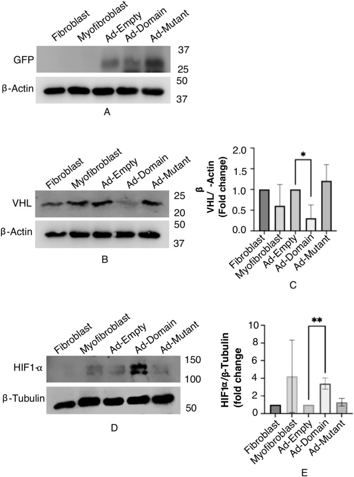
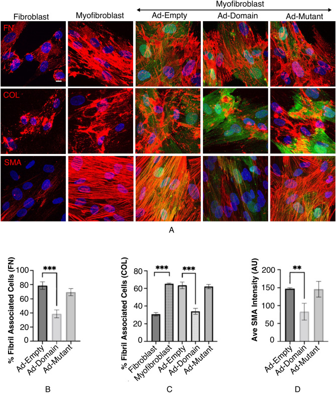
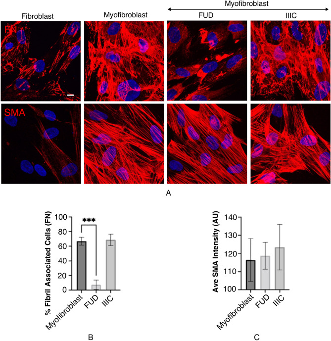
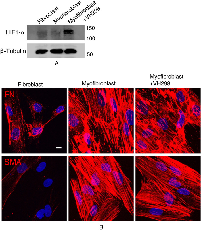
Idiopathic pulmonary fibrosis (IPF) is a fatal disease defined by a progressive decline in lung function due to scarring and accumulation of extracellular matrix (ECM) proteins. The SOCS (Suppressor Of Cytokine Signaling) domain is a 40 amino acid conserved domain known to form a functional ubiquitin ligase complex targeting the Von Hippel Lindau (VHL) protein for proteasomal degradation. Here we show that the SOCS conserved domain operates as a molecular tool, to disrupt collagen and fibronectin fibrils in the ECM associated with fibrotic lung myofibroblasts. Our results demonstrate that fibroblasts differentiated using TGFβ, followed by transduction with the SOCS domain, exhibit significantly reduced levels of the contractile myofibroblast-marker, α-SMA. Furthermore, in support of its role to retard differentiation, we find that lung fibroblasts expressing the SOCS domain present with significantly reduced levels of α-SMA and fibrillar fibronectin after differentiation with TGFβ. We show that adenoviral delivery of the SOCS domain in the fibrotic phase of experimental lung fibrosis in mice, significantly reduces collagen accumulation in disease lungs. These data underscore a novel function for the SOCS domain and its potential in ameliorating pathologic matrix deposition in lung fibroblasts and experimental lung fibrosis.

Keywords: ECM; Fibroblast; Fibronectin; Myofibroblast; SOCS domain.


Conflict of interest statement

Competing interests: The authors declare no competing interests.

Figures

Fig. 1
Fig. 1
SOCS domain degrades VHL protein in myofibroblasts. (A,B,D) IMR90 lung fibroblasts were untreated or differentiated with 5 ng/mL TGFβ for 48 h. Differentiated myofibroblasts were then transduced with Ad-Empty 100 MOI, Ad-Domain 600 MOI or Ad-Mutant 600 MOI for ~ 36 h. Cells were lysed in SDS lysis buffer and immunoblotted for SOCS-GFP fusion protein, VHL and HIF1-⍺. β-Actin and β-Tubulin were used as the loading controls. (C,E) Quantification of VHL and HIF1-⍺ normalized to their respective loading controls, on immunoblots are represented as averages of three independent trials. Statistical significance between fibroblasts and myofibroblasts and Ad-Empty and Ad-Domain or Ad-Mutant P-values were determined using Student’s t-test.
Fig. 2
Fig. 2
SOCS domain fragments FN matrix and reduces levels of SMA protein in myofibroblasts. (A) IMR90 lung fibroblasts were untreated or differentiated with 5 ng/mL TGFβ for 48 h. Differentiated myofibroblasts were then transduced with Ad-Empty (100 MOI), Ad-Domain (600 MOI) or Ad-Mutant (600 MOI) for ~ 36 h and immunostained for FN (red), COL (red) or SMA (red) and nuclear stain DAPI (blue). Myofibroblasts transduced with Ad-Empty (100 MOI), Ad-Domain (600 MOI) or Ad-Mutant (600 MOI) appear green (GFP reporter). Scale bar = 10 µm. (B,C) GFP reporter-expressing transduced cells as in (A) were quantified for percent of fibronectin fibril- containing cells and collagen fibril-containing cells respectively. Bar graphs are averages of three independent experiments. Student t-test was used to calculate P values. (D) Transduced cells expressing the GFP reporter as in (A) were used to quantify the SMA intensity values. Intensities shown in the graph are averages of three independent experiments. Student’s t-test was used to calculate P values.
Fig. 3
Fig. 3
SOCS domain reduces differentiation of fibroblasts to myofibroblasts. (A) IMR90 lung fibroblasts were untreated or transduced with Ad-Empty (100 MOI), Ad-Domain (600 MOI) or Ad-Mutant (600 MOI) for ~ 36 h. Cells were then differentiated with 5 ng/mL TGFβ for 48 h. Fibroblasts, myofibroblasts (untransduced) and transduced myofibroblasts were immunostained for FN (red) or SMA (red) and nuclear stain DAPI (blue). Myofibroblasts transduced with Ad-Empty (100 MOI), Ad-Domain (600 MOI) or Ad-Mutant (600 MOI) appear green (GFP reporter). Scale bar = 10 µm. (B) Transduced cells as in (A) were quantified for percent of fibronectin fibril- containing cells and represented as bar graphs. Only cells that expressed the GFP reporter were included in the analysis. (C) Transduced cells expressing the GFP reporter were used to quantify the SMA intensity values. Intensities shown in the graph are averages of three independent experiments. Student’s t-test was used to calculate P values.
Fig. 4
Fig. 4
FN fibril inhibition does not decrease SMA intensity in myofibroblasts. (A) IMR90 lung fibroblasts were untreated or differentiated with 5 ng/mL TGFβ. Differentiated myofibroblasts were then treated with 500 nM FUD or 500 nM IIIC-11C for 24 h in addition to 5 ng/mL TGFβ. Cells were immunostained for FN (red) or SMA (red) and nuclear stain, DAPI (blue). Scale bar = 10 µm. (B) Myofibroblasts and myofibroblasts treated with FUD or IIIC were quantified for fibronectin fibril- containing cells or (C) used to quantify the SMA intensity values. Bar graphs are averages of three independent trials. Student’s t-test was used to determine P values.
Fig. 5
Fig. 5
VHL protein inhibition does not decrease SMA intensity in myofibroblasts. (A) IMR90 lung fibroblasts were untreated or differentiated with 5 ng/mL TGFβ for 48 h. Differentiated myofibroblasts were then treated with 100 µM VH298 for 1 h. Cells were then lysed in SDS buffer and immunoblotted for HIF1-⍺. β-Tubulin was used as the loading control. (B) Fibroblasts, myofibroblasts and myofibroblasts treated as in (A) were immunostained for FN (red) or SMA (red) and nuclear stain, DAPI (blue). Scale bar = 10 µm.
Fig. 6
Fig. 6
Intranasal delivery of Adenoviral-GFP is localized to lung tissue and SOCS domain delivery significantly reduces collagen deposition in fibrotic lung. (A) C57BL/6 mice were intranasally administered vehicle (phosphate buffered saline) and adenoviral-GFP on Day 0 at 108 PFU and 109 PFU as depicted. Lung tissue was collected on Day 5 and Formalin Fixed paraffin embedded (FFPE) lung tissue was subjected to immunohistochemistry (IHC) staining using GFP-DAB antibody. Increased transduction of Ad-GFP at 109 PFU was observed compared to transduction at 108 PFU 5 days following intranasal delivery of Ad-GFP and vehicle control (n = 3). Magnification = 20x. Scale bar = 10 μm. (B) FFPE lung tissue sections analyzed for GFP expression. Top row depicts GFP-DAB staining and bottom row shows nuclei (blue). (C) Positive stain intensity from the bottom row in FIG. 6B, assigned using the scores: 1 + yellow, 2 + orange and 3 + red was used to quantify extent and intensity of GFP represented using the histoscore (H-score). Bar graph represents mean ± SD, n = 3. Statistical significance between vehicle (phosphate buffered saline) versus transduced and P-values (*P < 0.05, ***P < 0.001) are depicted. (D) C57BL/6 mice were administered bleomycin 3.07 units/kg by oropharyngeal aspiration on D0 and intranasally administered adenovirus on D9. Lung lobes collected on D21 was quantified for collagen deposits (hydroxyproline assay). Disease vs SOCS domain *p < 0.05. n = 6 mice per group.

Update of

Similar articles

Cited by

References

    1. Flaherty, K. R. et al. Radiological versus histological diagnosis in UIP and NSIP: survival implications. Thorax58, 143–148 (2003). - PMC - PubMed
    1. Monaghan, H. et al. Prognostic implications of histologic patterns in multiple surgical lung biopsies from patients with idiopathic interstitial pneumonias. Chest125, 522–526 (2004). - PubMed
    1. Perez, A., Rogers, R. M. & Dauber, J. H. The prognosis of idiopathic pulmonary fibrosis. Am. J. Respir. Cell Mol. Biol.29, S19-26 (2003). - PubMed
    1. Bethany, B. M. et al. Animal models of fibrotic lung disease. Am. J. Respir. Cell Mol. Biol.49, 167–179. 10.1165/rcmb.2013-0094TR (2013). - PMC - PubMed
    1. Raghu, G., Weycker, D., Edelsberg, J., Bradford, W. Z. & Oster, G. Incidence and prevalence of idiopathic pulmonary fibrosis. Am. J. Respir. Crit. Care Med.174, 810–816. 10.1164/rccm.200602-163OC (2006). - PubMed

MeSH terms

LinkOut - more resources