

Encapsulation of Beauveria bassiana conidia as a new strategy for the biological control of Aedes aegypti larvae
- PMID: 39738305
- PMCID: PMC11685528
- DOI: 10.1038/s41598-024-83036-9
Encapsulation of Beauveria bassiana conidia as a new strategy for the biological control of Aedes aegypti larvae
Abstract
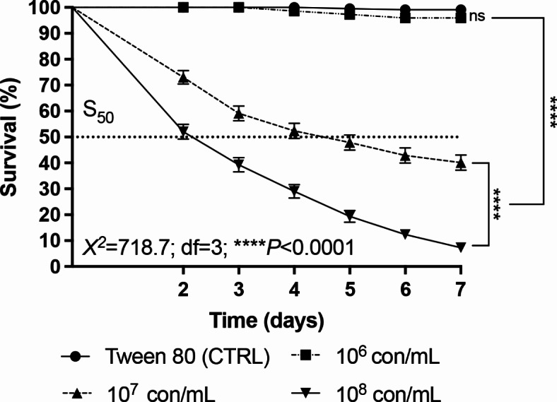
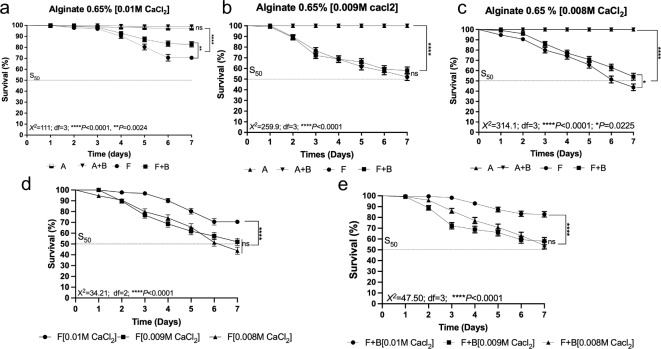
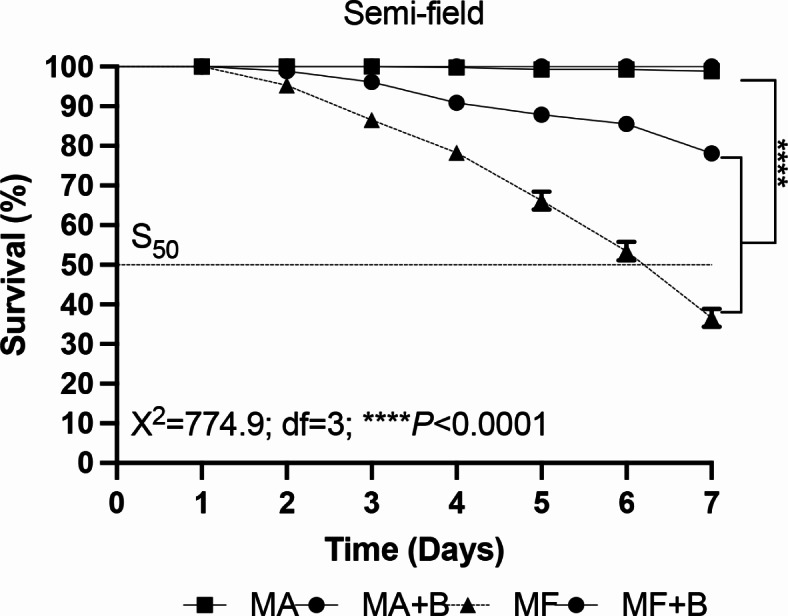
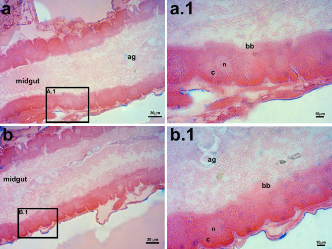
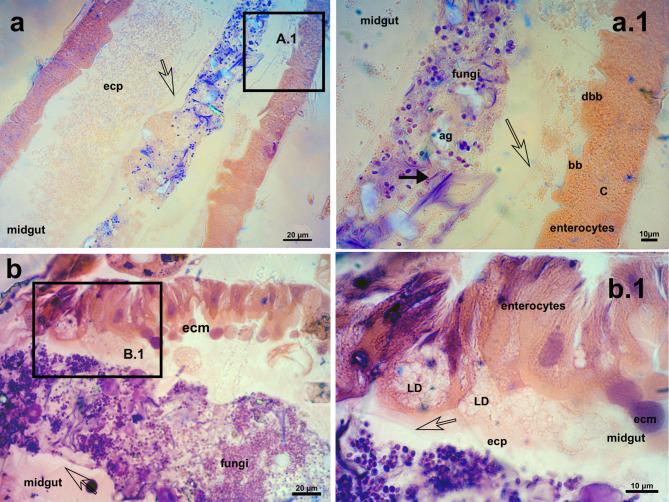
The virulence of encapsulated fungal conidia against Aedes aegypti larvae was investigated. Molecular studies confirmed that the fungal isolate used here was Beauveria bassiana. Different conidial concentrations were tested. A concentration of 1 × 10⁸ conidia mL- 1 was the most effective, resulting in 7% larval survival after 7 days. Next, alginate capsules (0.65%) containing conidia were prepared with different densities of calcium chloride (0.01 M, 0.009 M, and 0.008 M CaCl₂) and tested against larvae. Furthermore, groups of capsules were prepared with bird diet to act as an attractant. All capsule densities tested reduced larval survival (ranging from 22 to 67%). However, capsules with 0.008 M CaCl₂ were the most effective. Furthermore, fungus-only capsules were more efficient when compared to those containing bird diet. Laboratory and semi-field bioassays were conducted using mixtures of capsules with different densities. In the laboratory, survival ranged from 26 to 53%, whereas in semi-field conditions, 35%, and 80% survival was observed for groups exposed to fungus-only capsules or capsules containing diet, respectively. Histopathological studies of larvae exposed to capsules showed the presence of the fungus in the digestive tract and visible damage to enterocytes. These findings offer new insights into the biological control of Ae. aegypti larvae.
Keywords: alginate; biocontrol; capsules; dengue; enterocytes; entomopathogenic fungi.
© 2024. The Author(s).
Conflict of interest statement
Declarations. Competing interests: The authors declare no competing interests.
Figures
Similar articles
-
Expression of Bacillus thuringiensis toxin Cyt2Ba in the entomopathogenic fungus Beauveria bassiana increases its virulence towards Aedes mosquitoes.PLoS Negl Trop Dis. 2019 Jul 15;13(7):e0007590. doi: 10.1371/journal.pntd.0007590. eCollection 2019 Jul. PLoS Negl Trop Dis. 2019. PMID: 31306427 Free PMC article.
-
Recombinant Beauveria bassiana expressing Bacillus thuringiensis toxin Cyt1Aa: a promising approach for enhancing Aedes mosquito control.Microbiol Spectr. 2024 Jul 2;12(7):e0379223. doi: 10.1128/spectrum.03792-23. Epub 2024 May 29. Microbiol Spectr. 2024. PMID: 38809029 Free PMC article.
-
Larvicidal activity, route of interaction and ultrastructural changes in Aedes aegypti exposed to entomopathogenic fungi.Acta Trop. 2021 Jan;213:105732. doi: 10.1016/j.actatropica.2020.105732. Epub 2020 Nov 12. Acta Trop. 2021. PMID: 33188750
-
Beauveria bassiana interacts with gut and hemocytes to manipulate Aedes aegypti immunity.Parasit Vectors. 2023 Jan 17;16(1):17. doi: 10.1186/s13071-023-05655-x. Parasit Vectors. 2023. PMID: 36650591 Free PMC article.
-
Neem oil increases the efficiency of the entomopathogenic fungus Metarhizium anisopliae for the control of Aedes aegypti (Diptera: Culicidae) larvae.Parasit Vectors. 2015 Dec 30;8:669. doi: 10.1186/s13071-015-1280-9. Parasit Vectors. 2015. PMID: 26715150 Free PMC article.
References
-
- Kallás, E. G. et al. Live, attenuated, tetravalent butantan-dengue vaccine in children and adults. N Engl. J. Med.390 (5), 397–408. 10.1056/NEJMoa2301790 (2024). - PubMed
-
- Wilder-Smith, A. Dengue vaccine development: challenges and prospects. Curr. Opin. Infect. Dis.35 (5), 390–396. 10.1097/QCO.0000000000000871 (2022). - PubMed
-
- Smith, L. B., Kasai, S. & Scott, J. G. Pyrethroid resistance in Aedes aegypti and Aedes albopictus: important mosquito vectors of human diseases. Pestic Biochem. Phys.133, 1–12. 10.1016/j.pestbp.2016.03.005 (2016). - PubMed
Publication types
MeSH terms
Grants and funding
LinkOut - more resources
Full Text Sources
