. 2024 Dec 30;14(1):31901.
doi: 10.1038/s41598-024-83198-6.

Biocompatible ionized air alleviates rat osteoarthritis by modulating polarization from M1 to M2 macrophages

Affiliations

Biocompatible ionized air alleviates rat osteoarthritis by modulating polarization from M1 to M2 macrophages

Haoran Yu et al. Sci Rep. .

Abstract

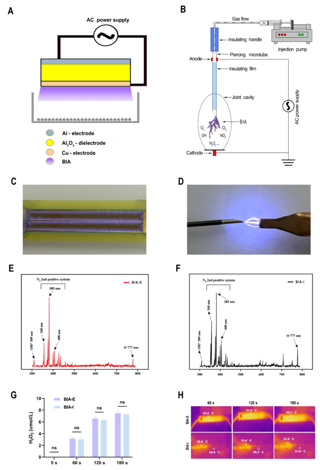
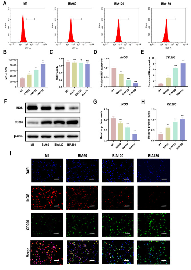
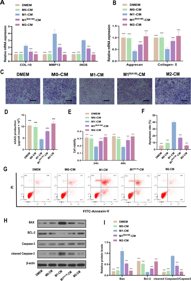
The imbalance in the proportion of M1/M2 macrophage polarization is a crucial contributor to the persistent progression of osteoarthritis (OA). This study aimed to evaluate the effects of low-dose biocompatible ionized air (BIA) on macrophage polarization and its subsequent chondroprotective effects, thereby validating the potential of BIA in slowing the progression of OA. In vitro experiments demonstrated that BIA modulates the polarization of M1 macrophages toward the M2 phenotype via the ROS-mediated STAT6 pathway. This shift reduces the expression of pro-inflammatory mediators while increasing the expression of anti-inflammatory mediators and pro-chondrogenic factors, leading to an improved microenvironment surrounding chondrocytes. The direct benefits of this improved microenvironment include enhanced chondrocyte viability, inhibition of apoptosis, and reduced degradation of the extracellular matrix. In vivo studies in rats showed that BIA inhibited M1 macrophage infiltration in the synovium, upregulated the proportion of M2 macrophages, alleviated cartilage degeneration, and delayed OA progression. This gas-based regulatory strategy may open new avenues for the treatment of OA.

Keywords: Biocompatible ionized air; Macrophages; Osteoarthritis.


Conflict of interest statement

Competing interests: The authors declare no conflicts of interest.

Figures

Fig. 1
Fig. 1
Schematic of BIA devices and plasma physicochemical parameters. (A) Operating schematic of BIA-E device. (B) Operating schematic of BIA-I device. (C) Physical discharge diagram of BIA-E device. (D) Physical discharge diagram of BIA-I device. (E)(F) Emission spectra of BIA-E and BIA-I devices between 300 and 800 nm. (G) Comparison of H2O2 levels generated by BIA-E and BIA-I devices at different operating times. (H) Working temperature of BIA-I and BIA-E devices over time. ‘ns’ indicates no significant difference between the two groups.
Fig. 2
Fig. 2
The effect of BIA on ROS levels, cellular viability, and polarization of M1 macrophages. (A), (B) DCFH-DA assay to evaluate BIA-induced changes in intracellular ROS levels in M1 macrophages. (C) CCK-8 assay to evaluate the effect of different BIA intervention durations on M1 macrophage viability. (D)–(E) qRT-PCR assay to evaluate gene expression levels of macrophage polarization markers (iNOS for M1 macrophages and CD206 for M2 macrophages) in M1 macrophages after different durations of BIA treatment. (F)–(H) Western blot to detect protein expression levels of macrophage polarization markers in M1 macrophages after different durations of BIA treatment. (I) Immunofluorescence assay to display representative fluorescence images of iNOS and CD206 in M1 macrophages after different durations of BIA treatment, with DAPI staining cell nuclei, scale bar = 50 μm. N = 3. *Significant difference to the M1 group: ***p < 0.001. Abbreviations: BIA: Biocompatible ionized air; ROS: reactive oxygen species; DCFH-DA: 2',7'- dichlorofluorescin diacetate assay; CCK-8: Cell Counting Kit-8; qRT-PCR: quantitative reverse transcription polymerase chain reaction; iNOS: inducible nitric oxide synthase; CD206: Cluster of Differentiation 206; DAPI: 4',6-diamidino-2-phenylindole.
Fig. 3
Fig. 3
BIA regulates M1 macrophage polarization to M2 macrophages through the ROS-mediated STAT6 pathway. (A) Western blot analysis of p-STAT6, iNOS, and CD206 expression levels in macrophages from different groups after BIA intervention. (B) Quantitative analysis of p-STAT6, iNOS, and CD206 expression levels in macrophages. (C) Immunofluorescence assay to visualize representative fluorescence images of p-STAT6 and corresponding macrophage polarization markers iNOS and CD206 in M1 macrophages after different intervention conditions, with DAPI staining cell nuclei. Scale bar = 20 μm. N = 3. *Significant difference compared to the M1 group: *p < 0.05, **p < 0.01, ***p < 0.001. #Significant difference compared to the M1BIA180 group: ##p < 0.01, ###p < 0.001. &Significant difference compared to the M1BIA180 group: &&p < 0.01, &&&p < 0.001. Abbreviations: BIA: Biocompatible ionized air; ROS: reactive oxygen species; STAT6: Signal transducer and activator of transcription 6; qRT-PCR: quantitative reverse transcription polymerase chain reaction; iNOS: inducible nitric oxide synthase; CD206: Cluster of Differentiation 206; DAPI: 4',6-diamidino-2-phenylindole; NAC: N-Acetyl-L-cysteine; LEF: Leflunomide.
Fig. 4
Fig. 4
Functional changes of M1 macrophages into M2 macrophages induced by BIA. (A)–(C) ELISA evaluation of pro-inflammatory cytokines IL-1β, IL-6, and TNF-α expression levels in M1 macrophages after BIA intervention. (D)–(F) ELISA evaluation of anti-inflammatory cytokines IL-10, TGF-β, and Arg-1 expression levels in M1 macrophages after BIA intervention. (G)–(P) Expression levels of pro-chondrogenic factors IGF1, IGF2, TGF-β1, TGF-β2, and TGF-β3 in M1 macrophages after BIA intervention. N = 3. *Significant difference compared to the M1 group: **p < 0.01, ***p < 0.001. Abbreviations: BIA: Biocompatible ionized air; IL-1β: Interleukin-1β; IL-6: Interleukin-6; TNF-α: Tumor necrosis factor α; IL-10: Interleukin-10; TGF-β: Transforming growth factor β; Arg-1: Arginase-1; IGF1: Insulin-like growth factor; TGF-β1: Transforming growth factor β1.
Fig. 5
Fig. 5
Different macrophage-CM co-culture with chondrocytes. (A) qRT-PCR evaluation of COL-10, MMP13, and iNOS expression levels in chondrocytes after 7 days of co-culture with different macrophage CM. (B) qRT-PCR evaluation of Aggrecan and Collagen II expression levels in chondrocytes after 7 days of co-culture with different macrophage CM. (C) Toluidine blue staining to assess GAG synthesis in chondrocytes after 7 days of co-culture with different macrophage CM. Scale bar = 100 μm. (D) Quantitative analysis of sGAG secretion in chondrocytes after 7 days of culture with different macrophage CM. (E) CCK-8 assay to evaluate chondrocyte viability at 24 h and 48 h after culture in different macrophage CM. (F)–(G) Quantitative analysis of chondrocyte apoptosis rate using flow cytometry and V-FITC/PI apoptosis assay after culture in different macrophage CM. (H)–(I) Quantitative analysis of apoptosis-related protein expression levels (BAX, BCL-2, Caspase-3, and cleaved Caspase-3) using Western blotting. N = 3. *Significant difference compared to the M1-CM group: ***p < 0.001. Abbreviations: BIA: Biocompatible ionized air; CM: conditioned media; DMEM: Dulbecco’s modified eagle medium; COL-10: Collagen-10; MMP13: Matrix metalloproteinase 13; iNOS: inducible nitric oxide synthase; GAG: Glycosaminoglycan; BAX: BCL-2-associated X protein; BCL-2: B-cell lymphoma 2.
Fig. 6
Fig. 6
In vivo effect of BIA treatment on rat knee OA. (A) Experimental design and schematic illustration of the rat knee OA model and evaluation of BIA’s protective effects. (B)–(E) Immunofluorescent staining of M1 (iNOS-positive) and M2 (CD206-positive) macrophages in the synovium. DAPI stains cell nuclei. Scale bar = 20 μm. (F) Hematoxylin and Eosin (H&E) staining. Scale bar = 50 μm. (G) Safranin and Fast Green (S&F) staining. Scale bar = 50 μm. (H) Three-dimensional micro-CT images (scale bar = 5 mm) and sagittal views of the knee joint (scale bar = 2 mm). (I)–(N) Quantitative analysis of M1/M2 macrophages, OARSI scores, BV, BV/TV, and Tb.N. N = 3. *Significant difference compared to the ACLT group: *p < 0.05, **p < 0.01, ***p < 0.001. &Significant difference compared to the Sham group: &&&p < 0.001. Abbreviations: BIA: Biocompatible ionized air; OA: Osteoarthritis; iNOS: inducible nitric oxide synthase; CD206: Cluster of Differentiation 206; ACLT: Anterior cruciate ligament transection; DAPI: 4',6-diamidino-2-phenylindole; H&E: Hematoxylin and eosin Staining; S&F: Safranin fast green staining; OARSI: Osteoarthritis research society international; BV: Bone volume; TV: Tissue volume; Tb.N: Trabecular number.
Fig. 7
Fig. 7
Further staining evaluation of BIA treatment on rat OA. (A) Immunohistochemical staining of MMP-13. Scale bar = 50 μm. (B) Immunohistochemical staining of Collagen II. Scale bar = 50 μm. (C) Immunohistochemical staining of Aggrecan. Scale bar = 50 μm. (D)–(F) Quantitative analysis of MMP-13, Collagen II, and Aggrecan. N = 3. *Significant difference compared to the ACLT group: *p < 0.05, **p < 0.01, ***p < 0.001. &Significant difference compared to the Sham group: &&&p < 0.001. Abbreviations: BIA: Biocompatible ionized air; OA: Osteoarthritis; ACLT: Anterior cruciate ligament transection; TUNEL: Terminal deoxynucleotidyl transferase dUTP; MMP-13: Matrix metalloproteinase 13.
Fig. 8
Fig. 8
Mechanisms of BIA in Alleviating Osteoarthritis.

Similar articles

References

    1. Hunter, D. J. & Bierma-Zeinstra, S. Osteoarthritis. Lancet393, 1745–1759. 10.1016/s0140-6736(19)30417-9 (2019). - PubMed
    1. Katz, J. N., Arant, K. R. & Loeser, R. F. Diagnosis and treatment of hip and knee osteoarthritis: A review. JAMA325, 568–578. 10.1001/jama.2020.22171 (2021). - PMC - PubMed
    1. Sun, W. et al. XJB-5-131 protects chondrocytes from ferroptosis to alleviate osteoarthritis progression via restoring Pebp1 expression. J. Orthopaedic Transl.44, 114–124. 10.1016/j.jot.2023.12.005 (2024). - PMC - PubMed
    1. Hu, Y. et al. Quercetin alleviates rat osteoarthritis by inhibiting inflammation and apoptosis of chondrocytes, modulating synovial macrophages polarization to M2 macrophages. Free Radic. Biol. Med.145, 146–160. 10.1016/j.freeradbiomed.2019.09.024 (2019). - PubMed
    1. Zhang, J., Rong, Y., Luo, C. & Cui, W. Bone marrow mesenchymal stem cell-derived exosomes prevent osteoarthritis by regulating synovial macrophage polarization. Aging12, 25138–25152. 10.18632/aging.104110 (2020). - PMC - PubMed

Publication types

Substances