

Systematic analysis of anatomy virtual reality (VR) apps for advanced education and further applications
- PMID: 39738416
- PMCID: PMC11685998
- DOI: 10.1038/s41598-024-82945-z
Systematic analysis of anatomy virtual reality (VR) apps for advanced education and further applications
Abstract
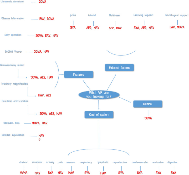
Virtual Reality (VR) technology enables users to immerse themselves in computer-generated environments, providing experiences that are otherwise difficult to attain in real life. VR has expanded from gaming into diverse fields, even the medical sector. In medical education, VR is mainly employed for anatomy and surgical practice, enhancing the learning experience by offering three-dimensional visualization and interaction with human structures. This article compares various VR anatomical applications, including "VEDAVI VR Human Anatomy", "Sharecare YOU Anatomy", "Everyday Anatomy VR", "3D Organon VR Anatomy", "Anatomy Explorer 2020", and "Human Anatomy VR". The comparison focuses on several aspects: the functions of each application, the accuracy of anatomical descriptions of the models, the depiction of movement and functionality of human body parts, the expression of disease, and other practical considerations such as tutorials, convenience of operation and observation, pricing, and user-friendliness. This study aims to serve as a guide for practical use, recommending the most suitable VR applications for educational purposes in the medical field and providing suggestions for improvement based on the analysis.
Keywords: Advanced education; Anatomy lecture; Hybrid class; Medical education; Virtual reality.
© 2024. The Author(s).
Conflict of interest statement
Competing interests: The authors declare no competing interests.
Figures


Similar articles
-
Creating 3D models from Radiologic Images for Virtual Reality Medical Education Modules.J Med Syst. 2019 May 3;43(6):166. doi: 10.1007/s10916-019-1308-3. J Med Syst. 2019. PMID: 31053902
-
Is virtual reality enhancing dental anatomy education? A systematic review and meta-analysis.BMC Med Educ. 2024 Nov 29;24(1):1395. doi: 10.1186/s12909-024-06233-0. BMC Med Educ. 2024. PMID: 39614238 Free PMC article.
-
Enhancing medical anatomy education with the integration of virtual reality into traditional lab settings.Clin Anat. 2025 Apr;38(3):249-268. doi: 10.1002/ca.24213. Epub 2024 Sep 8. Clin Anat. 2025. PMID: 39245875
-
Twelve tips for teaching in virtual reality.Med Teach. 2024 Apr;46(4):495-499. doi: 10.1080/0142159X.2023.2285396. Epub 2023 Nov 25. Med Teach. 2024. PMID: 38006603
-
A new dimension in medical education: Virtual reality in anatomy during COVID-19 pandemic.Clin Anat. 2023 Oct;36(7):1007-1015. doi: 10.1002/ca.24098. Epub 2023 Jul 24. Clin Anat. 2023. PMID: 37485993 Review.
Cited by
-
Enhancing anatomy education with virtual reality: integrating three-dimensional models for improved learning efficiency and student satisfaction.Front Med (Lausanne). 2025 Jun 4;12:1555053. doi: 10.3389/fmed.2025.1555053. eCollection 2025. Front Med (Lausanne). 2025. PMID: 40534689 Free PMC article. Review.
-
Research on the Implementation Status and Optimization Path of "Internet + Nursing Service" from the Perspective of Grade Hospital Difference: A Multi-Dimensional Empirical Study Based on Online Nurses in Quzhou City.J Multidiscip Healthc. 2025 Aug 13;18:4989-4999. doi: 10.2147/JMDH.S532942. eCollection 2025. J Multidiscip Healthc. 2025. PMID: 40827216 Free PMC article.
References
-
- Garcia Barrios, A., Cisneros Gimeno, A. I., Garza García, M. C., Lamiquiz Moneo, I. & Whyte Orozco, J. Online teaching alternative in human anatomy. Anat.1, 86–90 (2022).
-
- Larson, R. C. & Strehle, G. P. Edu-tech: What’sa president to do. Technol. Enhanc. Learn. 21–59 (2002).
-
- Shwab, K. The Fourth Industrial Revolution: What It Means, How to Respond (World Economic Forum, 2016).
-
- Trelease, R. B. From chalkboard, slides, and paper to e-learning: How computing technologies have transformed anatomical sciences education. Anat. Sci. Educ.9, 583–602 (2016). - PubMed
-
- Simonetti, V. et al. Effectiveness of virtual reality in the management of paediatric anxiety during the peri-operative period: A systematic review and meta-analysis. Int. J. Nurs. Stud.125, 104115 (2022). - PubMed
Publication types
MeSH terms
LinkOut - more resources
Full Text Sources
Medical
