. 2024 Dec 30;14(1):32087.
doi: 10.1038/s41598-024-83815-4.

PPAR-γ agonist mitigates intestinal barrier dysfunction and inflammation induced by Clostridioides difficile SlpA in vitro

Affiliations

PPAR-γ agonist mitigates intestinal barrier dysfunction and inflammation induced by Clostridioides difficile SlpA in vitro

Maryam Noori et al. Sci Rep. .

Abstract

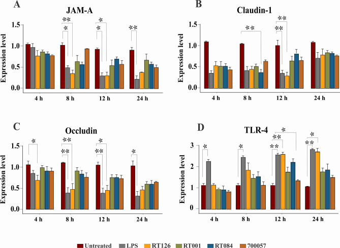
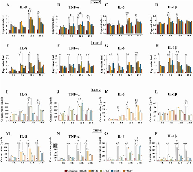
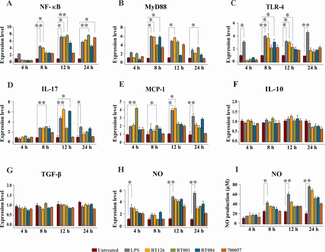
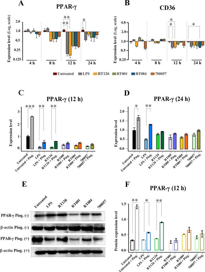
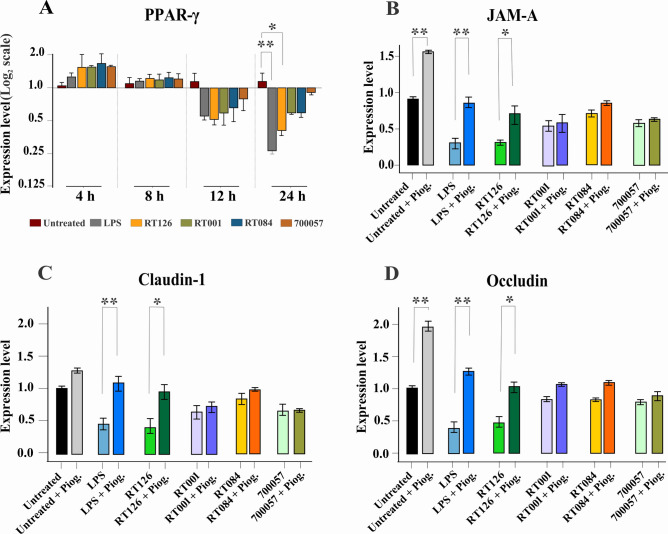
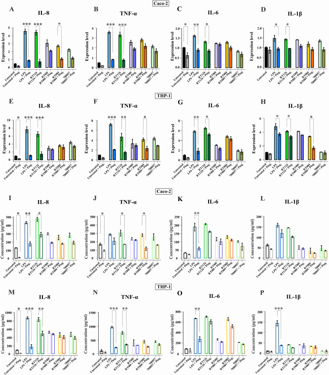
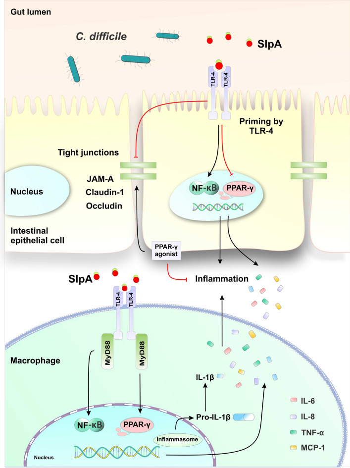
Clostridioides difficile is the leading cause of healthcare- and antibiotic-associated diarrhea. Surface layer protein A (SlpA), an essential component of the bacterium's outermost layer, contributes to colonization and inflammation. The peroxisome proliferator-activated receptor gamma (PPAR-γ) has been demonstrated to improve intestinal integrity and prevent inflammation in host cells. Here, we investigated the role of PPAR-γ in SlpA-mediated inflammation in Caco-2 cells and THP-1 derived macrophages. The extraction of SlpA was carried out for three toxigenic C. difficile clinical strains (RT126, RT001, RT084) and a non-toxigenic strain (ATCC 700057). The gene expression of tight junction (TJ) proteins and inflammatory markers was determined using RT-qPCR. The production of proinflammatory cytokines and nitric oxide was measured by ELISA and Griss reaction, respectively. Western blotting was performed to detect PPAR-γ level before and after adding its agonist, pioglitazone. SlpA of C. difficile strains enhanced the expression of TLR-4, NF-κB, MyD88, IL-17, MCP-1, IL-8, IL-6, TNF-α, IL-1β, whilst the gene expression level of JAM-A, claudin-1, occludin, PPAR-γ and its receptor (CD36) was decreased in both Caco-2 cells and THP-1 derived macrophages. Moreover, pioglitazone caused a notable elevation in the expression level of PPAR-γ, only following treatment with RT126 SlpA. Besides, pioglitazone pretreatment improved TJ impairment in Caco-2 cells and attenuated proinflammatory cytokine expression in both SlpA-treated cell lines. SlpA can attenuate PPAR-γ expression, trigger TJ disruption, and stimulate inflammatory response in host cells. Notably, these events could be reversed by pretreatment of cells with PPAR-γ agonist. Further experiments are required to corroborate the present findings.

Keywords: Clostridioides difficile; Caco-2 cells; Peroxisome proliferator-activated receptor gamma; Pioglitazone; Surface layer protein A; THP-1 derived macrophages.


Conflict of interest statement

Competing interests: The authors declare no competing interests.

Figures

Fig. 1
Fig. 1
The effect of C. difficile SlpA on tight junction-associated markers and TLR-4. Relative gene expression of (A) JAM-A, (B) claudin-1, (C) occludin, (D) TLR-4 in Caco-2 cells upon treatment with SlpA of C. difficile (RT126, RT001, RT084) and SlpA of C. difficile ATCC 700057 determined by RT-qPCR. Adjusted P values less than 0.05 were considered statistically significant; *P < 0.05, **P < 0.01, and ***P < 0.001.
Fig. 2
Fig. 2
The effect of C. difficile SlpA on proinflammatory cytokines expression and production. Relative gene expression of (A) IL-8, (B) TNF-α, (C) IL-6, and (D) IL-1β in Caco-2 cells, and (E) IL-8, (F) TNF-α, (G) IL-6, and (H) IL-1β in THP-1 derived macrophages upon treatment with SlpA of C. difficile (RT126, RT001, RT084) and SlpA of C. difficile ATCC 700057 determined by RT-qPCR. Production of (I) IL-8, (J) TNF-α, (K) IL-6, and (L) IL-1β in Caco-2 cells, and (M) IL-8, (N) TNF-α, (O) IL-6, and (P) IL-1β in THP-1 derived macrophages upon treatment with SlpA purified from C. difficile (RT126, RT001, RT084) and SlpA of C. difficile ATCC 700057 measured by ELISA. Adjusted P values less than 0.05 were considered statistically significant; *P < 0.05, **P < 0.01, and ***P < 0.001.
Fig. 3
Fig. 3
Effect of C. difficile SlpA on the expression of inflammatory-related markers. Relative expression of (A) NF-κB, (B) MyD88, (C) TLR-4, (D) IL-17, (E) MCP-1, (F) IL-10, (G) TGF-β, and (H) NO genes was determined by RT-qPCR assay, and concentration of (I) NO was measured by Griss reaction in THP-1 derived macrophages upon treatment with SlpA of C. difficile (RT126, RT001, RT084) and SlpA of C. difficile ATCC 700057. Adjusted P values less than 0.05 were considered statistically significant; *P < 0.05, **P < 0.01, and ***P < 0.001.
Fig. 4
Fig. 4
The role of PPAR-γ and its agonist, pioglitazone, in C. difficile SlpA-driven inflammation. Relative expression of (A) PPAR-γ and (B) CD36 genes was determined by RT-qPCR assay in THP-1 derived macrophages upon treatment with SlpA of C. difficile (RT126, RT001, RT084) and SlpA of C. difficile ATCC 700,057. Additionally, the relative expression of PPAR-γ was determined at (C) 12 h, and (D) 24 h of treatment with SlpA in the absence and presence of pioglitazone. THP-1 derived macrophages were also analyzed by using western blotting for the expression levels of PPAR-γ in the absence and presence of pioglitazone at 12 h of treatment (E, F). The optical densities of the bands were calculated by ImageJ software. Adjusted P values less than 0.05 were considered statistically significant; *P < 0.05, **P < 0.01, and ***P < 0.001. Piog., pioglitazone.
Fig. 5
Fig. 5
The effect of C. difficile SlpA on tight-junction associated markers in the absence and presence of pioglitazone in Caco-2 cells. Relative expression of (A) PPAR-γ, (B) JAM-A, (C) claudin-1, and (D) occludin genes was determined by RT-qPCR in Caco-2 cells after 12 h of treatment with SlpA of C. difficile (RT126, RT001, RT084) and SlpA of C. difficile ATCC 700057 in the presence and absence of pioglitazone. Adjusted P values less than 0.05 were considered statistically significant; *P < 0.05, **P < 0.01, and ***P < 0.001. Piog., pioglitazone.
Fig. 6
Fig. 6
The effect of C. difficile SlpA on proinflammatory cytokines expression and production in the absence and presence of pioglitazone. Relative gene expression of (A) IL-8, (B) TNF-α, (C) IL-6, and (D) IL-1β in Caco-2 cells, and (E) IL-8, (F) TNF-α, (G) IL-6, and (H) IL-1β in THP-1 derived macrophages upon 24 h of treatment with SlpA of C. difficile (RT126, RT001, RT084) and SlpA of C. difficile ATCC 700057 in the presence and absence of pioglitazone determined by RT-qPCR. Production of (I) IL-8, (J) TNF-α, (K) IL-6, and (L) IL-1β in Caco-2 cells, and (M) IL-8, (N) TNF-α, (O) IL-6, and (P) IL-1β in THP-1 derived macrophages upon 24 h of treatment with SlpA of C. difficile (RT126, RT001, RT084) and SlpA of C. difficile ATCC 700057 in the presence and absence of pioglitazone measured by ELISA. Adjusted P values less than 0.05 were considered statistically significant; *P < 0.05, **P < 0.01, and ***P < 0.001. Piog., pioglitazone.
Fig. 7
Fig. 7
Potential mechanism of C. difficile SlpA-mediated inflammation and intestinal barrier impairment. SlpA can disrupt tight junctions and subsequently may increase intestinal permeability in intestinal epithelial cells. It can also interact with TLR-4, leading to an increased level of proinflammatory mediators in the gut lumen. In addition, SlpA can downregulate the expression of PPAR-γ, thereby promoting inflammatory response by activation of TLR-4/NF-κB pathway. Pretreatment of intestinal epithelial cells with PPAR-γ agonist can mitigate SlpA-induced tight junction disruption. PPAR-γ agonist can also reduce SlpA-induced inflammation in both intestinal epithelial cells and macrophages. Furthermore, SlpA is able to enhance the production of IL-1β, potentially by processing pro-IL-1β into its active form through the activation of inflammasome pathway.

Similar articles

Cited by

References

    1. Czepiel, J. et al. Clostridium difficile infection: review. Eur. J. Clin. Microbiol. Infect. Dis.38, 1211–1221. 10.1007/s10096-019-03539-6 (2019). - PMC - PubMed
    1. De Roo, A. C. & Regenbogen, S. E. Clostridium difficile infection: an epidemiology update. Clin. Colon Rectal. Surg.33, 49–57. 10.1055/s-0040-1701229 (2020). - PMC - PubMed
    1. Cherny, K. E., Muscat, E. B., Reyna, M. E. & Kociolek, L. K. Clostridium innocuum: microbiological and clinical characteristics of a potential emerging pathogen. Anaerobe71, 102418. 10.1016/j.anaerobe.2021.102418 (2021). - PMC - PubMed
    1. Lee, H. Y. et al. Risk factors and outcomes of Clostridium difficile infection in hospitalized patients. Biomed. J.42, 99–106. 10.1016/j.bj.2018.12.002 (2019). - PMC - PubMed
    1. Chandrasekaran, R. & Lacy, D. B. The role of toxins in Clostridium difficile infection. FEMS Microbiol. Rev.41, 723–750. 10.1093/femsre/fux048 (2017). - PMC - PubMed

Publication types

LinkOut - more resources