. 2024 Dec 30;14(1):32068.
doi: 10.1038/s41598-024-83764-y.

Therapeutic effects of resveratrol and β-carotene on L-arginine-induced acute pancreatitis through oxidative stress and inflammatory pathways in rats

Affiliations

Therapeutic effects of resveratrol and β-carotene on L-arginine-induced acute pancreatitis through oxidative stress and inflammatory pathways in rats

Arzu Gezer et al. Sci Rep. .

Abstract

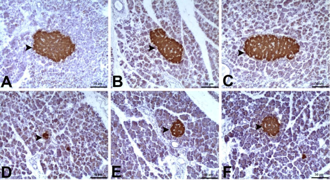
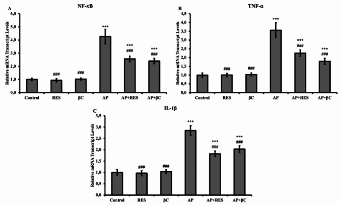
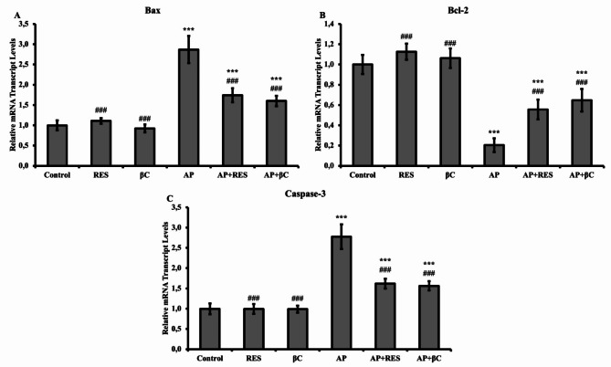
Acute pancreatitis (AP) is a severe inflammatory condition affecting the pancreas, often leading to systemic inflammation and organ dysfunction. This study evaluated the effects of resveratrol (RES) and β-carotene (βC) on L-arginine-induced AP in rats. Forty-eight male Sprague Dawley rats were divided into six groups: Control (C), RES (20 mg/kg), βC (50 mg/kg), AP, AP + RES, and AP + βC. The AP model was induced with 250 mg/100 g L-arginine intraperitoneally twice daily with a 1-h interval. The AP group showed significantly elevated oxidative stress (MDA) and reduced GSH levels (p < 0.001). Immunohistochemical (IHC) staining with anti-insulin antibody revealed reduced β + langerhans islet size in the AP group. qPCR analysis indicated significant upregulation of inflammatory genes NF-κB, TNF-α, and IL-1β (p < 0.001), and apoptotic genes Bax and Caspase-3, with downregulation of Bcl-2 (p < 0.001). RES and βC treatments significantly reduced MDA levels and increased GSH levels (p < 0.01 for both) compared to the AP group. The AP + RES and AP + βC groups exhibited preserved β + Langerhans islet size (p < 0.01), suppressed NF-κB, TNF-α, and IL-1β expression, reduced Bax and Caspase-3 levels, and increased Bcl-2 levels (p < 0.01). Histopathological findings supported these results. RES and βC confer significant effects against L-arginine-induced acute pancreatitis by reducing oxidative stress, preserving pancreatic islet integrity, suppressing inflammatory responses, and modulating apoptotic pathways. RES demonstrated a slightly superior efficacy in reducing inflammation and oxidative stress markers, suggesting it may be more effective in treating acute pancreatitis.

Keywords: Acute pancreatitis; Apoptosis; L-arginine; Oxidative stress; Resveratrol; İnflammation; β‐carotene.


Conflict of interest statement

Declarations. Competing interests: The authors declare no competing interests.

Figures

Fig. 1
Fig. 1
Histopathological examination of the pancreas in different groups: (A) Control group, (B) RES group, (C) βC group. Normal histological appearance. (D) AP group. Severe degenerative-necrotic changes in the cells of Langerhans islets (arrow) and acinar cells (arrowhead). (E) AP + RES group. Severe degenerative-necrotic changes in the cells of Langerhans islets (arrow) and moderate changes in acinar cells (arrowhead). (F) AP + βC group. Mild degenerative-necrotic changes in the cells of Langerhans islets (arrow) and acinar cells (arrowhead) (H&E staining, magnification ×40).
Fig. 2
Fig. 2
Levels of oxidative stress markers MDA and GSH in pancreatic tissues. Values are expressed as mean ± SD. Statistical significance: Control vs. others: *** p < 0.001; AP vs. others: ### p < 0.001.
Fig. 3
Fig. 3
Diameter and area of β + Langerhans islets (arrowhead) in different groups (immunohistochemical staining with anti-insulin antibody). (A) Control group, (B) RES group, (C) βC group. Normal histological appearance of β + Langerhans islets. (D) AP group. Group with the smallest β + Langerhans islets. (E) AP + RES group. Group with larger β + Langerhans islets. (F) AP + βC group. Group with the largest β + Langerhans islets (Immunohistochemistry, magnification × 20).
Fig. 4
Fig. 4
Relative mRNA transcript levels of inflammatory genes NF-κB, TNF-α, and IL-1β in pancreatic tissues. Statistical significance: Control vs. others: *** p < 0.001; AP vs. others: ### p < 0.001.
Fig. 5
Fig. 5
Relative mRNA transcript levels of apoptotic genes Bax and Caspase-3, and anti-apoptotic gene Bcl-2 in pancreatic tissues. Statistical significance: Control vs. others: *** p < 0.001; AP vs. others: ### p < 0.001.

Similar articles

Cited by

References

    1. Mederos, M. A., Reber, H. A. & Girgis, M. D. Acute pancreatitis: A review. JAMA325(4), 382–390 (2021). - PubMed
    1. Gagyi, E.-B. et al. Incidence of recurrent and chronic pancreatitis after acute pancreatitis: A systematic review and meta-analysis. Ther. Adv. Gastroenterol.17, 17562848241255304 (2024). - PMC - PubMed
    1. Iannuzzi, J. P. et al. Global incidence of acute pancreatitis is increasing over time: A systematic review and meta-analysis. Gastroenterology162(1), 122–134 (2022). - PubMed
    1. Szatmary, P. et al. Acute pancreatitis: Diagnosis and treatment. Drugs82(12), 1251–1276 (2022). - PMC - PubMed
    1. Pérez, S., Pereda, J., Sabater, L. & Sastre, J. Redox signaling in acute pancreatitis. Redox Biol.5, 1–14 (2015). - PMC - PubMed

MeSH terms