. 2024 Dec 30;14(1):31998.
doi: 10.1038/s41598-024-83644-5.

Investigating skeletal muscle micro-trauma with time-dependent diffusion and the random permeable barrier model

Affiliations

Investigating skeletal muscle micro-trauma with time-dependent diffusion and the random permeable barrier model

Susanne S Rauh et al. Sci Rep. .

Abstract

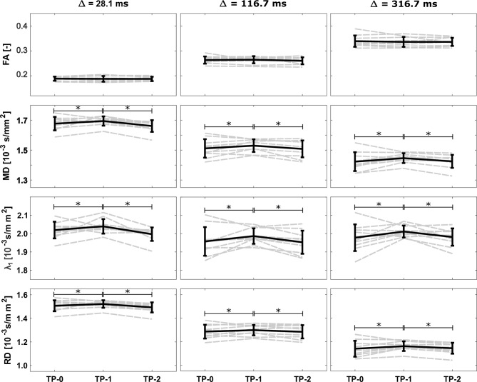
Repeated muscle micro-trauma may cause severe muscle damage. Diffusion tensor imaging (DTI) exhibits sensitivity to microstructural changes in skeletal muscle. We hypothesize that longer diffusion times enhance sensitivity to micro-trauma and that membrane permeability increases with micro-trauma. We obtained DTI scans of the thighs in ten male runners 1 week before (TP-0), 24-48 h after (TP-1), and 2 weeks after (TP-2) they completed a marathon. Diffusion times were 28.1, 116.7, and 316.7 ms. The random permeable barrier model (RPBM) was fitted to the radial diffusivities to obtain estimates for fiber diameter and membrane permeability. Hamstring and quadriceps muscles were manually segmented. A linear mixed model assessed variations across time points and diffusion times within the DTI and RPBM parameters and assessed sensitivity to micro-trauma by comparing %-changes in DTI parameters at TP-1 and TP-2 to TP-0. All DTI parameters except FA significantly changed between TP-0 and TP-1, and between TP-1 and TP-2. The %-change did not differ between diffusion times. The permeability increased at TP-1 and TP-2 compared to TP-0. In conclusion, longer diffusion times did not improve sensitivity to micro-trauma. The increased permeability post-marathon underscores the potential of RPBM-derived parameters as a biomarker for micro-trauma and muscle injuries.


Conflict of interest statement

Declarations. Competing interests: H.E.K. reports research support from Philips Healthcare during the conduct of the study, and trial support from ImagingDMD-UF outside the submitted work. All reimbursements for H.E.K. were received by the LUMC. No personal financial benefits were received. All other authors do not hold any competing interest.

Figures

Fig. 1
Fig. 1
Field of view and muscle segmentations. Left: Location chosen for the field of view centered at mid-thigh level, shown on a surface render plot of the upper legs. Right: Axial Dixon water image with segmentations drawn in the hamstring and quadriceps muscles of both legs. VL: vastus lateralis, VI: vastus intermedius, RF: rectus femoris, VM: vastus medialis, BFS: biceps femoris short head, BFL: biceps femoris long head, ST: semitendinosus, SM: semimembranosus.
Fig. 2
Fig. 2
Conventional DTI analysis. Mean fractional anisotropy (FA), mean diffusivity (MD), first diffusion tensor eigenvalue (λ1, axial diffusivity), and radial diffusivity (RD) parameters, averaged over all muscles, for each subject (n = 10, dashed gray lines) at all three time points (TP-0 to TP-2) and diffusion times (Δ). The mean and standard deviation over all subjects are shown in black. * = p < 0.001.
Fig. 3
Fig. 3
Random permeable barrier model (RPBM) analysis. Left: Axial (top, blue) and radial (bottom, orange) diffusion vs. diffusion time of the right semitendinosus (filled circles) and the right rectus femoris (open circles) at TP-0 for a representative subject. While the changes in axial diffusivity over diffusion time are subtle, the decrease in radial diffusivity with increasing diffusion time is more pronounced. The dashed lines show the RPBM fits to the radial diffusivity (black: semitendinosus, gray: rectus femoris). Right: Line plots of the fiber diameter, a, and the permeability, κ, averaged over all muscles, for each subject (n = 10, dashed gray lines). The mean and standard deviation over all subjects are overlaid in black. * p ≤ 0.008.
Fig. 4
Fig. 4
Percentage change in DTI parameters. The boxplots show the relative change between TP-1 and TP-0 for λ1, radial diffusivity (RD) and fractional anisotropy (FA) for each diffusion time. The individual datapoints for each subject, muscle and leg (left/right) are overlaid in blue (n = 160). The red line indicates no change between TP-1 and TP-0 (0%). Although the median values are similar for all diffusion times, some very high percentage changes are found in RD at the diffusion time of 316.7 ms.
Fig. 5
Fig. 5
Random permeable barrier model (RPBM) parameter maps. Maps of the fiber diameter (top) and permeability (bottom) of one subject are shown at all three time points (TP-0 – TP-2). Between the time points, no clear differences are visible in the maps. The excluded voxels that failied the quality control step of the radial diffusivity (RD) (RD(Δ = 28.1 ms) > RD(Δ = 116.7 ms) > RD(Δ = 316 ms)) are black in both maps, the fiber diameter and permeability.
Fig. 6
Fig. 6
Injury analysis. Mean diffusivity (MD) and radial diffusivity (RD) at all diffusion times (Δ) (a) and the RPBM parameters a and κ (b) of the injured muscles (n = 25, dashed gray lines) at all three time points (TP-0 to TP-2) are shown. The mean and standard deviation over all injuries are overlaid in black. * p ≤ 0.05.

Similar articles

Cited by

References

    1. Hyldahl, R. D. & Hubal, M. J. Lengthening our perspective: Morphological, cellular, and molecular responses to eccentric exercise. Muscle Nerve. 49, 155–170. 10.1002/mus.24077 (2014). - PubMed
    1. Fridén, J., Sjoestroem, M. & Ekblom, B. A morphological study of delayed muscle soreness. Experientia37, 506–507 (1981). - PubMed
    1. Clarkson, P. M. & Tremblay, I. Exercise-induced muscle damage, repair, and adaption in humans. J. Appl. Physiol.65, 1–6 (1988). - PubMed
    1. Butterfield, T. A. Eccentric exercise in vivo: Strain-induced muscle damage and adaptation in a stable system. Exerc. Sport Sci. Rev.38, 51–60. 10.1097/JES.0b013e3181d496eb (2010). - PubMed
    1. Reurink, G. et al. Magnetic resonance imaging in acute hamstring injury: Can we provide a return to play prognosis? Sport Med.45, 133–146. 10.1007/s40279-014-0243-1 (2014). - PubMed

Publication types

LinkOut - more resources