. 2024 Dec 30;14(1):32003.
doi: 10.1038/s41598-024-83578-y.

Micromorphological features of brown rotted wood revealed by broad argon ion beam milling

Affiliations

Micromorphological features of brown rotted wood revealed by broad argon ion beam milling

Rikako Tsukida et al. Sci Rep. .

Abstract

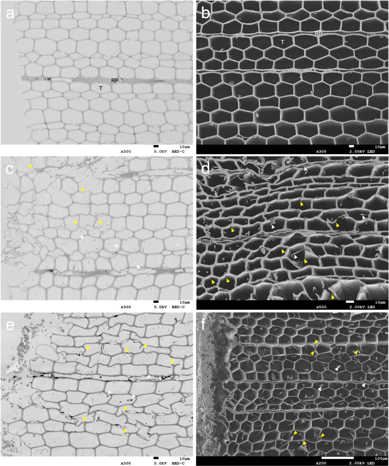
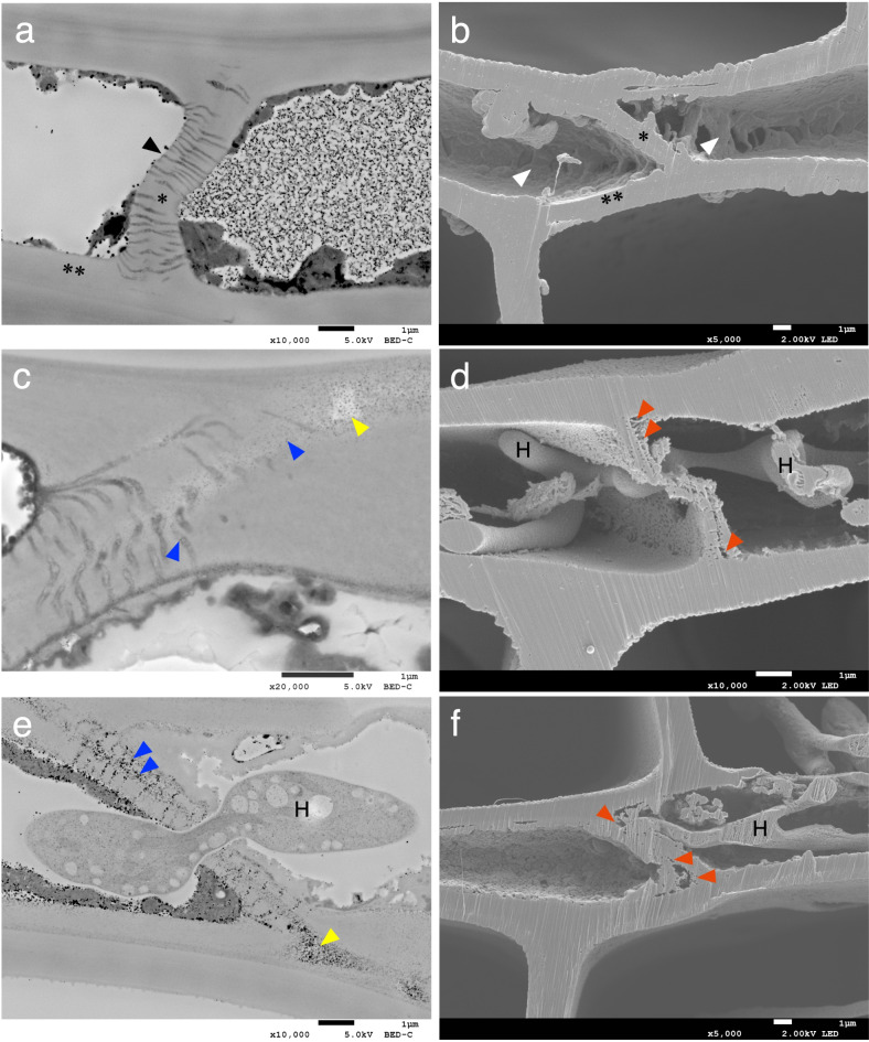
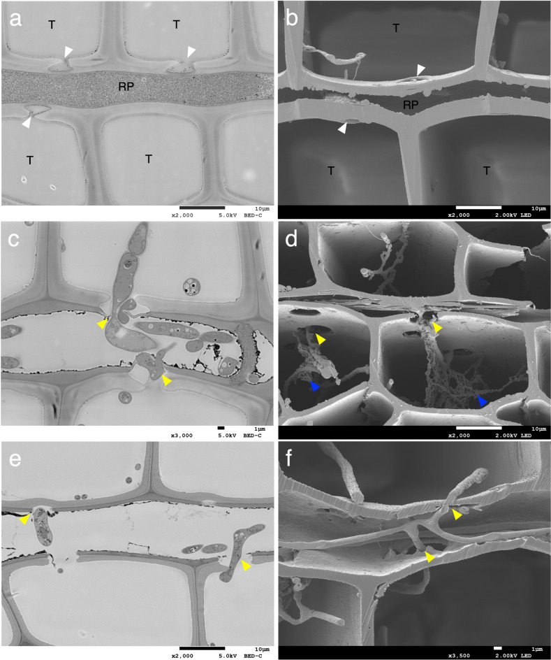
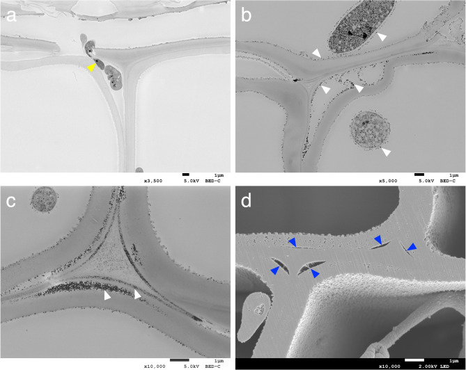
Brown rot fungi, the major decomposers in the boreal coniferous forests, cause a unique wood decay pattern but many aspects of brown rot decay mechanisms remain unclear. In this study, decayed wood samples were prepared by cultivation of the brown rot fungi Gloeophyllum trabeum and Coniophora puteana on Japanese coniferous wood of Cryptomeria japonica, and the cutting planes were prepared using broad ion beam (BIB) milling, which enables observation of intact wood, in addition to traditional microtome sections. Samples were observed using field-emission SEM revealing that areas inside the end walls of ray parenchyma cells were the first to be degraded. Osmium reaction precipitates were observed in the degraded regions, as well as in plasmodesmata. In the cell wall where ray parenchyma cells contacted with the tracheids, specific degradation of cross-field pits and hyphal elongation into this area was observed in degradation by both fungi. Other pit types were also degraded as noted in previous studies. Delamination between the S1 and S2 layers of tracheids, and cracks in the tracheid cell walls were observed. These findings provide new insights into the cell wall degradation mechanisms during the incipient stages of brown rot decay.

Keywords: Broad ion beam milling; Brown rot fungi; Ray parenchyma cells; Tracheids; Wood degradation.


Conflict of interest statement

Declarations. Competing interests: The authors declare no competing interests.

Figures

Fig. 1
Fig. 1
Low-magnification transverse section images: (a,b) undecayed wood, (c,d) G. trabeum 32 days, (e,f) C. puteana 12 days. Images a, c and e are sections prepared using an ultra-microtome. Images b, d and f are cross-sections obtained through BIB milling. White arrowheads indicate hyphae. Yellow arrowheads indicate cracks. RP = ray parenchyma; T = tracheid.
Fig. 2
Fig. 2
Observation of ray parenchyma cells: (a,b) undecayed wood, (c,d) G. trabeum 32 days, (e) C. puteana 32 days, (f) C. puteana 12 days. Images a, c and e are sections prepared using an ultra-microtome. Images b, d and f are cross-sections obtained through BIB milling. A black arrowhead indicates plasmodesmata. White arrowheads indicate cellular content in the ray parenchyma cells. Blue arrowheads indicate osmium-reaction precipitates (ORPs). Yellow arrowheads indicate the areas appeared to have greater transparency. Red arrowheads indicate degradation areas within the end wall, including areas where small pores, and coalesced pores were created as part of the decay process. H = hyphae; * = end walls; ** = the cell walls of ray parenchyma cells in contact with tracheids.
Fig. 3
Fig. 3
Image analysis of the end walls of ray parenchyma cells. (a) The size of ORPs in the decayed wood samples by G. trabeum 32 days and C. puteana 32 days. Statistical significance was calculated using a Welch’s t-test. *P < 0.05. (b) The size of the pores in the end walls of ray parenchyma cells in Fig. 2b,d,f, respectively. Pores larger than 400 nm were included in the next rank.
Fig. 4
Fig. 4
Observation of the cross-field pitting region. (a,b) undecayed wood. (c,d) G. trabeum 32 days. (e,f) C. puteana 12 days. Images a, c and e are sections prepared using an ultra-microtome. Images b, d and f are cross-sections obtained through BIB milling. White arrowheads indicate cross-field pits. Yellow arrowheads indicate hyphal elongation through cross-field pits. Blue arrowheads indicate characteristic materials around hyphae. RP = ray parenchyma; T = tracheid.
Fig. 5
Fig. 5
Delamination between the S1 and S2 layers in tracheid cell walls: (a) G. trabeum 12 days, (b,c) C. puteana 16 days, (d) G. trabeum 32 days. Images a–c are sections prepared using an ultra-microtome. Image d shows cross-sections obtained through BIB milling. A yellow arrowhead indicates hyphae elongation into the delamination area. White arrowheads indicate ORPs. Blue arrowheads indicate delamination.

Similar articles

References

    1. Highley, T. L. Changes in chemical components of hardwood and softwood by brown-rot fungi. Mater. Organ.22, 39–46 (1987).
    1. Kirk, T. K. & Highley, T. L. Quantitative changes in structural components of conifer woods during decay by white- and brown-rot fungi. Phytopathology63, 1338–1342 (1973).
    1. Kaffenberger, J. T. & Schilling, J. S. Comparing lignocellulose physiochemistry after decomposition by brown rot fungi with distinct evolutionary origins. Environ. Microbiol.17, 4885–4897 (2015). - PubMed
    1. Winandy, J. E. & Morrell, J. J. Relationship between incipient decay, strength, and chemical composition of Douglas-fir heartwood. Wood Fiber Sci.25, 278–288 (1993).
    1. Kleman-Leyer, K., Agosin, E., Conner, A. H. & Kirk, T. K. Changes in molecular size distribution of cellulose during attack by white rot and brown rot fungi. Appl. Environ. Microbiol.58, 1266–1270 (1992). - PMC - PubMed

Publication types

LinkOut - more resources