

MiR-363-3p induces tamoxifen resistance in breast cancer cells through PTEN modulation
- PMID: 39738797
- PMCID: PMC11685982
- DOI: 10.1038/s41598-024-83938-8
MiR-363-3p induces tamoxifen resistance in breast cancer cells through PTEN modulation
Abstract
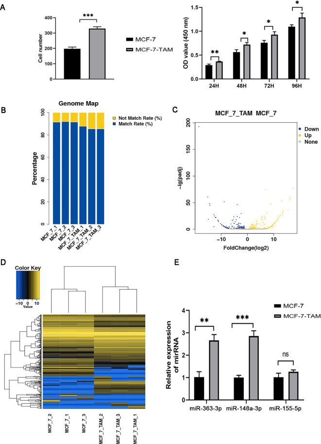
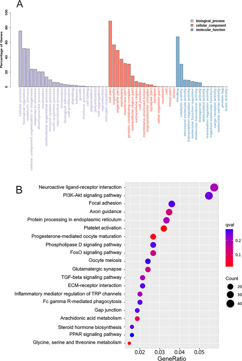
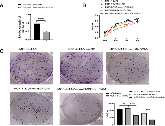
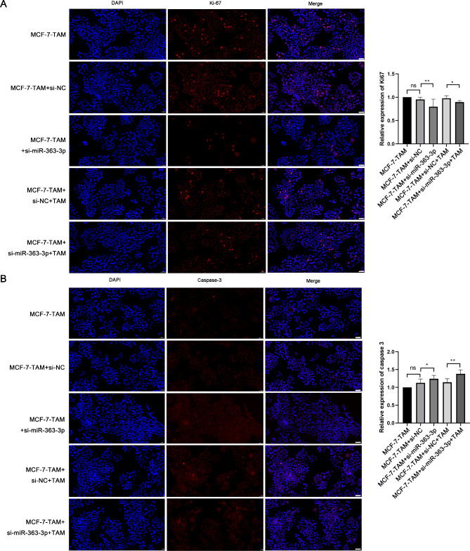
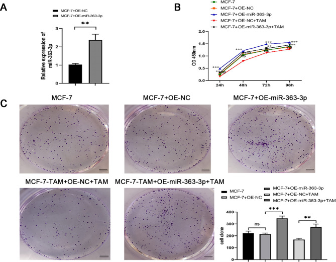
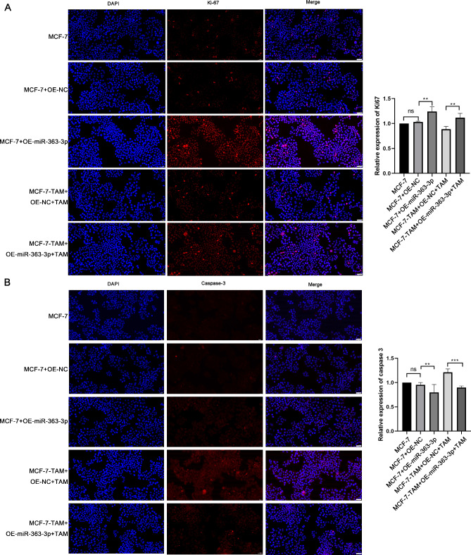
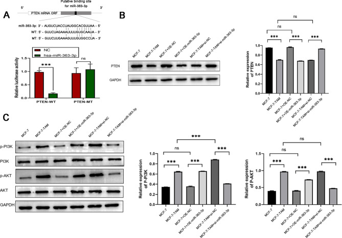
Nowadays, the investigation for overcoming tamoxifen (TAM) resistance is confronting a considerable challenge. Therefore, immediate attention is required to elucidate the mechanism underlying TAM resistance in breast cancer. This research primarily aimed to define how miRNA-363-3p facilitates resistance to TAM in breast cancer. High-throughput miRNA sequencing was performed using RNAs prepared from breast cancer MCF-7 cells and TAM-resistant MCF-7 cells (MCF-7-TAM). An increase in miRNA-363-3p levels was observed in MCF-7-TAM cells. In MCF-7 cells, miRNA-363-3p directly targeted and negatively regulated phosphatase and tensin homolog (PTEN). Reduction of miRNA-363-3p retarded cell growth and accelerated cell apoptosis, thereby enhancing the sensitivity of TAM. Moreover, analysis using the Kyoto Encyclopedia of Genes and Genomes (KEGG) pathway showed significant enrichment of target genes within the phosphoinositide-3-kinase (PI3K)/protein kinase B (AKT) signaling pathway. Ultimately, miR-363-3p decreased the responsiveness of breast cancer cells to TAM by targeting and suppressing PTEN through a mechanism associated with the PI3K-Akt pathway. Therefore, these results suggest that miR-363-3p-dependent PTEN expression contributes to the mechanisms underlying breast cancer endocrine resistance.
Keywords: Breast cancer; Hsa-miR-363-3p; PI3K/AKT signaling pathway; PTEN; TAM resistance.
© 2024. The Author(s).
Conflict of interest statement
Declarations. Competing interests: The authors declare no competing interests. Ethics approval: This study was approved by the Ethics Committee of Shandong Provincial Hospital Affiliated to Shandong First Medical University (SMYX: NO. 2019-092). Consent to participate: Not applicable. Consent to publish: Not applicable.
Figures
Similar articles
-
Silencing of MicroRNA-21 confers the sensitivity to tamoxifen and fulvestrant by enhancing autophagic cell death through inhibition of the PI3K-AKT-mTOR pathway in breast cancer cells.Biomed Pharmacother. 2016 Feb;77:37-44. doi: 10.1016/j.biopha.2015.11.005. Epub 2015 Dec 12. Biomed Pharmacother. 2016. PMID: 26796263
-
MicroRNA-29a contributes to drug-resistance of breast cancer cells to adriamycin through PTEN/AKT/GSK3β signaling pathway.Gene. 2016 Nov 15;593(1):84-90. doi: 10.1016/j.gene.2016.08.016. Epub 2016 Aug 11. Gene. 2016. PMID: 27523474
-
Keratinocyte growth factor (KGF) regulates estrogen receptor-alpha (ER-alpha) expression and cell apoptosis via phosphatidylinositol 3-kinase (PI3K)/Akt pathway in human breast cancer cells.Anticancer Res. 2009 Aug;29(8):3195-205. Anticancer Res. 2009. PMID: 19661335
-
MicroRNA dynamics, PTEN/PI3K/AKT signaling, and their relationship to breast cancer: prospects for pharmaceuticals and natural product application.Breast Cancer Res Treat. 2025 Feb;209(3):467-485. doi: 10.1007/s10549-024-07600-7. Epub 2025 Jan 10. Breast Cancer Res Treat. 2025. PMID: 39792295 Review.
-
Targeting signal transduction pathways to eliminate chemotherapeutic drug resistance and cancer stem cells.Adv Enzyme Regul. 2010;50(1):285-307. doi: 10.1016/j.advenzreg.2009.10.016. Epub 2009 Nov 4. Adv Enzyme Regul. 2010. PMID: 19895837 Free PMC article. Review. No abstract available.
Cited by
-
Self-control moderates the impacts of physical activity on the sleep quality of university students.Sci Rep. 2025 Feb 3;15(1):4040. doi: 10.1038/s41598-025-88700-2. Sci Rep. 2025. PMID: 39900789 Free PMC article.
References
-
- Sung, H. et al. Global Cancer statistics 2020: GLOBOCAN estimates of incidence and mortality worldwide for 36 cancers in 185 countries. CA Cancer J. Clin.71, 209–249 (2021). - PubMed
-
- Hutchinson, L. Breast cancer: Challenges, controversies, breakthroughs. Nat. Rev. Clin. Oncol.7, 669–670 (2010). - PubMed
-
- Anastasiadi, Z., Lianos, G. D., Ignatiadou, E., Harissis, H. V. & Mitsis, M. Breast cancer in young women: An overview. Updates Surg.69, 313–317 (2017). - PubMed
-
- Zhang, X. et al. Monopolar spindle 1 contributes to tamoxifen resistance in breast cancer through phosphorylation of estrogen receptor α. Breast Cancer Res. Treat.202, 595–606 (2023). - PubMed
Publication types
MeSH terms
Substances
Grants and funding
LinkOut - more resources
Full Text Sources
Medical
Research Materials
