

Case study of a neuroendocrine tumor of uncertain origin: single-cell transcriptomics unravels potential primary location
- PMID: 39738894
- PMCID: PMC11688255
- DOI: 10.1007/s00432-024-06071-z
Case study of a neuroendocrine tumor of uncertain origin: single-cell transcriptomics unravels potential primary location
Abstract
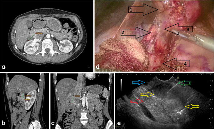
Purpose: Determining the primary origin of non-organ-confined neuroendocrine tumors (NETs) for accurate diagnosis and management. Neuroendocrine tumors are rare neoplasms with diverse clinical behaviors. Determining their primary origin remains challenging in cases of non-organ-confined NETs. This study explores the histogenesis of a retroperitoneal, non-functional NET localized between the duodenum and pancreatic head, utilizing advanced molecular diagnostics to elucidate its probable primary source.
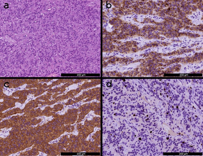
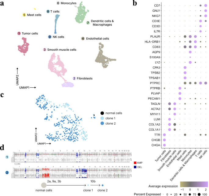
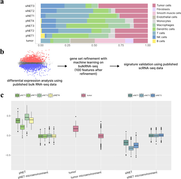
Methods: Initial diagnostic methods, including imaging and histopathology, failed to resolve the tumor's origin. The tumor was subjected to single-cell RNA sequencing (scRNA-seq) and whole exome sequencing (WES). Publicly available transcriptomic datasets from pancreatic and small intestine NETs were used to develop and validate a molecular gene signature for tissue-of-origin identification.
Results: The gene signature distinguished pancreatic and small intestine NETs with high accuracy. The tumor cells presented a molecular profile consistent with a pancreatic origin, likely derived from ectopic pancreatic tissue.
Conclusions: This case demonstrates the value of integrating scRNA-seq and WES for the molecular characterization of complex NETs. Identifying the tumor's pancreatic origin informed a targeted management approach, avoiding unnecessary systemic treatment and underscoring the potential of single-cell approaches in personalized oncology.
Keywords: Duodenal NET; Ectopic pancreatic tissue; Neuroendocrine tumor; Non-organ-confined NET; Pancreatic NET; Single-cell RNA‐seq.
© 2024. The Author(s).
Conflict of interest statement
Declarations. Competing interests: The authors declare no competing interests.
Figures
References
-
- Alvarez MJ, Subramaniam PS, Tang LH, Grunn A, Aburi M, Rieckhof G, Komissarova EV, Hagan EA, Bodei L, Clemons PA et al (2018) A precision oncology approach to the pharmacological targeting of mechanistic dependencies in neuroendocrine tumors. Nat Genet 50:979–989. 10.1038/s41588-018-0138-4 - DOI - PMC - PubMed
-
- Barkas N, Petukhov V, Kharchenko P, Steiger S, Rydbirk R, Biederstedt E (2021) Pagoda2: Single Cell Analysis and Differential Expression. R package version 1.0.12
Publication types
MeSH terms
Grants and funding
LinkOut - more resources
Full Text Sources
Medical
Molecular Biology Databases
