. 2025 Feb;12(8):e2411602.
doi: 10.1002/advs.202411602. Epub 2024 Dec 31.

Chirality-Induced Hydroxyapatite Manipulates Enantioselective Bone-Implant Interactions Toward Ameliorative Osteoporotic Osseointegration

Affiliations

Chirality-Induced Hydroxyapatite Manipulates Enantioselective Bone-Implant Interactions Toward Ameliorative Osteoporotic Osseointegration

Liang Yang et al. Adv Sci (Weinh). 2025 Feb.

Abstract

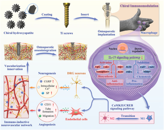
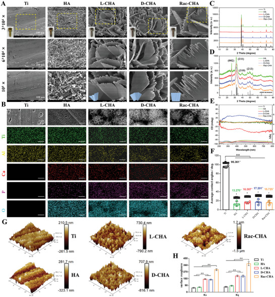
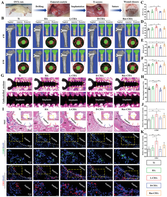
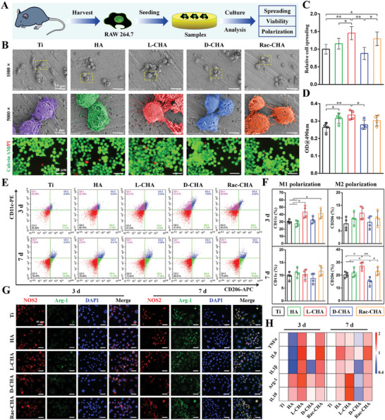
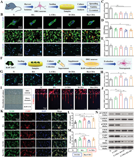
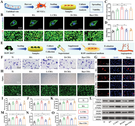
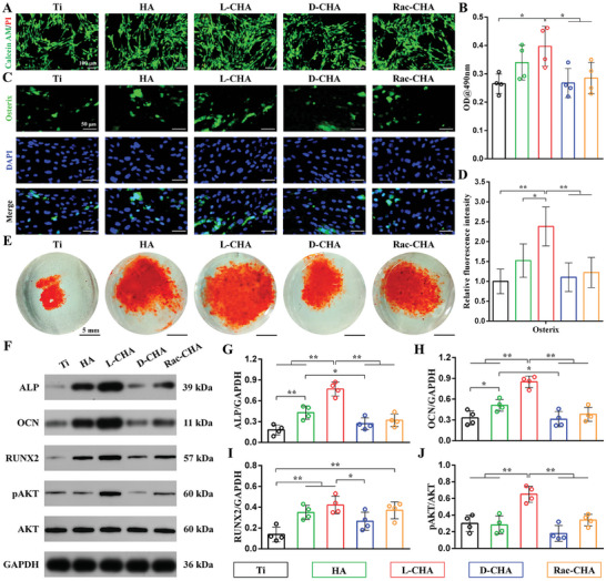
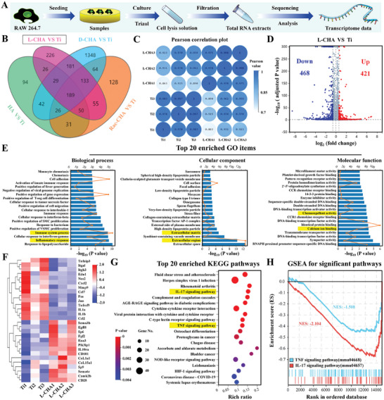
Inspired by the fundamental attribute of chirality in nature, chiral-engineered biomaterials now represent a groundbreaking frontier in biomedical fields. However, the integration of chirality within inorganic materials remains a critical challenge and developments of chirality-induced bionic bone implants are still in infancy. In this view, novel chiral hydroxyapatite (CHA) coated Ti alloys are successfully synthesized by a sophisticated chiral molecule-induced self-assembly method for the first time. The obtained samples are characterized by stereospecific L-/D-/Rac-chiral hierarchical morphology, nanotopography rough surfaces, improved hydrophilicity, and bioactivity. Following implantation into rat femoral condyle defects, the distinct stereospecific chiral hierarchical structures exhibit highly enantioselective bone-implants interactions, wherein the left-handed chirality of L-CHA strongly promotes osteoporotic osseointegration and vice versa for right-handed chirality of D-CHA. Consistently, in vitro assays further validate the superior enantiomer-dependent osteoporotic osseointegration ability of L-CHA, mainly by manipulating desired immunomodulation coupled with enhanced neurogenesis, angiogenesis, and osteogenesis. Moreover, as analyzed by transcriptomic RNA-seq, a new discovery of down-regulated IL-17 signaling pathway is considered predominately responsible for the desired immunomodulation ability of L-CHA. These results provide new insights into biological multifunctionality and mechanism underlying L-chirality's roles for bone healing, thus may inspiring developments of new generation of chiral biomaterials.

Keywords: chirality; enantioselectivity; hydroxyapatite; immunomodulation; osteoporotic osseointegration.


Conflict of interest statement

The authors declare no conflict of interest.

Figures

Figure 1
Figure 1
Schematic illustration of L‐CHA coated Ti which manipulates desired immunomodulation coupled with enhanced neurogenesis and angiogenesis toward ameliorative osteoporotic osseointegration.
Figure 2
Figure 2
Physicochemical properties characterizations of CHA coated Ti. A) Macroscopical view and representative SEM images of samples at various magnifications. The areas marked by yellow box are magnified for observation. B) SEM‐EDS mapping of samples including Ti, Al, Ca, P, and O elements. C,D) XRD patterns of (C) CHA‐coated Ti and (D) synthesized powder of CHA coating. E) Chirality of samples determined by UV–vis and CD spectra. F) Surface hydrophilicity of samples quantified by contact angles. G,H) Surface roughness of samples examined by AFM and analyzed based on Ra and Rq parameters. n = 4. ** p < 0.01.
Figure 3
Figure 3
Evaluation of implants osseointegration by micro‐CT and histological analysis. A) Implantation procedures of materials into femoral condyle defects of OVX rats. B) Representative sagittal, anteroposterior axial, and median transverse images of implants reconstructed by 3D micro‐CT. The median transverse area was marked by red lines and magnified for observation (green for implanted Ti screws and red for bone formation). C–F) Quantitative analysis of micro‐CT data in terms of (C,D) BV/TV and (E,F) BMD. G,H) Undecalcified sagittal sections of samples and quantification of BIC at 8 weeks post‐implantation. I) Median transverse sections of samples stained by H&E (first row), CGRP (second and third row), and CD31 (fourth and fifth row) immunohistofluorescence at 4 weeks post‐implantation. J,K) Quantitative fluorescence intensity of (J) CGRP and (K) CD31. The areas marked by yellow box are magnified for observation; Black and white dashed lines indicate bone‐implant interface; Red arrows mark peri‐implant bone formation; Blue arrows mark fibrous tissues; HB represents host bone. n = 4. * p < 0.05, ** p < 0.01.
Figure 4
Figure 4
Macrophage responses to CHA‐coated Ti samples. A) Procedures of cell seeding on samples and biological examinations. B) RAW cells adhesion morphology and viability detected by SEM (first and second row) and live/dead staining (third row) respectively. The areas marked by yellow box are magnified and rendered for observation. C) RAW cells spreading quantified by aspect ratios. (D) RAW cells viability determined by MTT. E,F) RAW cells polarization quantified by CD11c (M1) and CD206 (M2) expressions using flow cytometry. G) RAW cells polarization evaluated by co‐staining of NOS2/Arg‐1 (M1/M2) immunofluorescence. H) Heatmap of pro‐inflammatory (TNFα, IL6, IL1β) and anti‐inflammatory (Arg‐1, IL10) gene expressions of RAW cells determined by RT‐PCR. n = 4. * p < 0.05, ** p < 0.01.
Figure 5
Figure 5
Cellular activities of DRG neurons cultured on CHA coated Ti and the immuno‐inductive neurogenesis evaluation of samples. A) Procedures of cell seeding on samples and biological examinations. B) The adhesion morphology, viability, and bioactivity of DRG neurons detected by cell actin cytoskeleton staining (first row), live/dead staining (second row), and Ca2+ fluorescence probe (third row) respectively. C) Cell spreading quantified by aspect ratios. D) Viability of DRG neurons determined by MTT. E) Quantification of intracellular Ca2+ based on fluorescence intensity of Ca2+ probe. F) In vitro immuno‐inductive neurogenesis of DRG neurons stimulated by RAW conditioned medium. G,H) Immuno‐inductive intracellular Ca2+ expressions examined by Ca2+ fluorescence probe. I,J) Immuno‐inductive DRG neurons axonal growth determined by microfluidics assays. White dashed lines indicate microgroove barriers (500 µm) of the microfluidic device. K) Immuno‐inductive CGRP and NF200 expressions detected by immunofluorescence co‐staining. L,M) Immuno‐inductive (L) CGRP and (M) SP expressions quantified by ELISA. N) Western blot analysis of immuno‐inductive CGRP and SP expressions in L‐CHA supplemented with/without KN‐93 or KG‐501. n = 4. * p < 0.05, ** p < 0.01.
Figure 6
Figure 6
Cellular activities of HUVECs cultured on CHA‐coated Ti and the immuno‐inductive angiogenesis evaluation of samples. A) Procedures of cell seeding on samples and biological examinations. B) The adhesion morphology and viability of HUVECs detected by cell actin cytoskeleton staining (first row) and live/dead staining (second row) respectively. C) Cell spreading quantified by aspect ratios. D) Viability of HUVECs determined by MTT. E) In vitro immuno‐inductive angiogenesis of HUVECs stimulated by RAW‐conditioned medium. F,I) Immuno‐inductive migration of HUVECs examined by transwell assays. G) Immuno‐inductive CD31 expressions detected by immunofluorescence. H) Immuno‐inductive tube formation detected via ordinary microscope (first row) and calcein AM fluorescence (second row). J,K) Quantification of (J) branch points number and (K) tube length based on tube formation images. L) Immuno‐inductive angiogenesis‐related proteins (VEGF, eNOS, FGF2) expressions detected by western blot. M–O) Quantification of (M) VEGF, (N) eNOS, and (O) FGF2 expressions based on western blot results. n = 4. * p < 0.05, ** p < 0.01.
Figure 7
Figure 7
Osteogenesis of BMSCs cultured on CHA‐coated Ti. A) Cell viability detected by live/dead staining for 3 days culture. B) Cell viability determined by MTT assays. C,D) Fluorescent quantitation of osterix expressions. E) ARS staining for 14 days culture. F) Osteogenesis‐related proteins (ALP, OCN, RUNX2, pAKT) expressions analyzed by western blot. G–J) Quantification of (G) ALP, (H) OCN, (I) RUNX2, and (J) pAKT expressions based on western blot results. n = 4. * p < 0.05, ** p < 0.01.
Figure 8
Figure 8
Mechanism of samples on modulating macrophage reactions analyzed by transcriptomic RNA‐seq. A) Procedures of RAW cells culture and subsequent RNA‐seq analysis. B) Venn diagram of differentially expressed genes (DEGs) amounts between CHA‐coated Ti and pristine Ti. C) Pearson correlation plot of L‐CHA and pristine Ti samples. D) Volcano plot of DEGs in L‐CHA versus pristine Ti. E) GO analysis of DEGs in L‐CHA versus pristine Ti. F) Heatmap of partial DEGs mainly involved in inflammation, angiogenesis, and neurogenesis. G) Top 20 enriched KEGG pathways based on DEGs of L‐CHA versus pristine Ti. H) GSEA analysis for significant pathways (IL‐17 and TNF signaling pathway) down‐regulated by L‐CHA versus pristine Ti. The yellow marks highlight interested GO items and KEGG pathways.

Similar articles

References

    1. Zhang X. D., Li Q. M., Wang Z. X., Zhou W., Zhang L. L., Liu Y. S., Xu Z., Li Z., Zhu C., Zhang X. Z., Front. Bioeng. Biotechnol. 2022, 10, 921092. - PMC - PubMed
    1. Jiang W., Hou F. S., Gu Y., Saiding Q., Bao P. P., Tang J. C., Wu L., Chen C. M., Shen C. L., Pereira C. L., Sarmento M., Sarmento B., Cui W. G., Chen L., Bioact. Mater. 2021, 12, 169 . - PMC - PubMed
    1. Gu H., Zhu Y. H., Yang J. W., Jiang R. X., Deng Y. W., Li A. S., Fang Y. J., Wu Q. J., Tu H. H., Chang H. S., Wen J., Jiang X. Q., Adv. Sci. 2023, 10, e2302136. - PMC - PubMed
    1. Zhao Y. N., Kang H. L., Wu X. P., Zhuang P. Z., Tu R., Goto T., Li F., Dai H. L., Adv. Healthcare Mater. 2023, 12, e2203099. - PubMed
    1. Zheng Y. Y., Gao A., Bai J. X., Liao Q., Wu Y. Z., Zhang W., Guan M., Tong L. P., Geng D. C., Zhao X., Chu P. K., Wang H. Y., Bioact. Mater. 2022, 14, 364. - PMC - PubMed

LinkOut - more resources