

Establishment of an oral burn model in streptozotocin-induced diabetic rats
- PMID: 39739075
- PMCID: PMC11685340
- DOI: 10.1186/s40902-024-00453-6
Establishment of an oral burn model in streptozotocin-induced diabetic rats
Abstract
Background: Oral ulcers are painful mucosal lesions prone to infection and inflammation. To evaluate the effectiveness of treatments, a suitable experimental animal model with an appropriate healing period is required. The aim of this study was to develop an animal model for oral ulcer research by comparing oral burn wounds of different sizes and locations in diabetic rats.
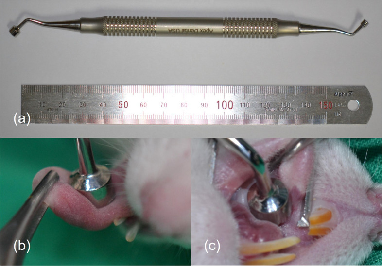
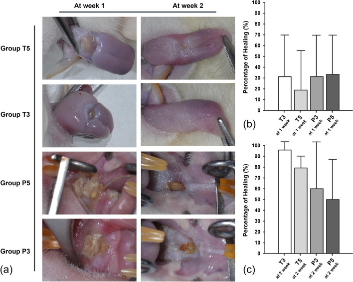
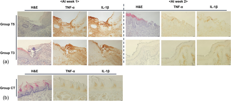
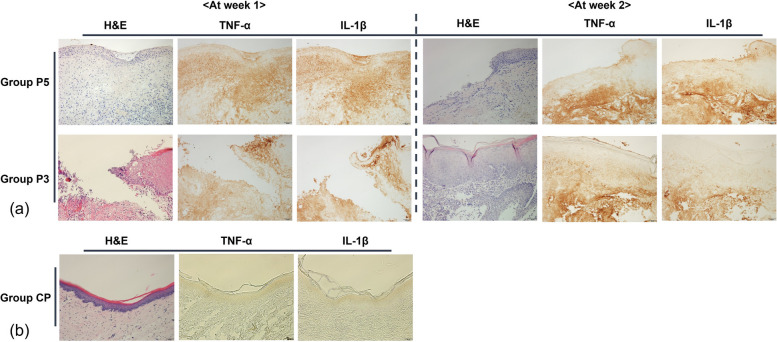
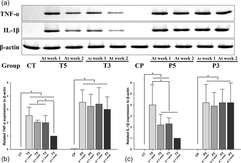
Methods: Forty-four male Sprague-Dawley rats with induced diabetes were divided into six groups based on burn wound location and size: T5 (n = 10, tongue 5 mm), T3 (n = 10, tongue 3 mm), P5 (n = 10, palate 5 mm), P3 (n = 10, palate 3 mm), CT (n = 2, control tongue), and CP (n = 2, control palate). The burn wounds were induced by applying a heated device (100-120 °C) for 3 s. At 1- and 2-weeks post-surgery, macroscopic examination, histological staining, immunohistochemistry, and Western blot analysis were performed to compare the healing progress.
Results: Healing progressed more rapidly in the second week than in the first for all groups, with burns on the tongue (Groups T5 and T3) showing more advanced healing compared to burns on the palate (Groups P5 and P3). By the second week, Group T3 was almost completely healed, while Group T5 had some remaining wounds. In contrast, Groups P5 and P3 showed minimal healing. This faster healing on the tongue was further supported by significantly lower expression levels of TNF-α and IL-1β and a reduction in ulcer size, particularly on the tongue compared to the palate.
Conclusion: A 3 mm or 5 mm burn wound on the tongue of diabetic rats can serve as a useful animal model for evaluating new treatments for wound healing, particularly up to the first week. However, for studies extending to the second week, the 5 mm burn wound model on the tongue might be more advantageous.
Keywords: Burns; Diabetes mellitus; Oral ulcer; Rat; Wound healing.
© 2024. The Author(s).
Conflict of interest statement
Declarations. Ethics approval and consent to participate: This study was approved by the Institutional Animal Care and Use Committee at Gangneung-Wonju National University (GWNU-2022-6). This study adheres to ARRIVE guidelines. Consent for publication: Not applicable. Competing interests: The authors declare that they have no competing interests.
Figures

Similar articles
-
[Analysis of differential gene expressions of inflammatory and repair-related factors in chronic refractory wounds in clinic].Zhonghua Shao Shang Za Zhi. 2019 Jan 20;35(1):18-24. doi: 10.3760/cma.j.issn.1009-2587.2019.01.005. Zhonghua Shao Shang Za Zhi. 2019. PMID: 30678397 Chinese.
-
The effect of dressing with fresh kiwifruit on burn wound healing.Surgery. 2010 Nov;148(5):963-8. doi: 10.1016/j.surg.2010.02.013. Epub 2010 Apr 8. Surgery. 2010. PMID: 20381106
-
[Effects and mechanism of astragalus polysaccharide on wound healing of deep partial-thickness burns in rats].Zhonghua Shao Shang Yu Chuang Mian Xiu Fu Za Zhi. 2023 Mar 20;39(3):256-263. doi: 10.3760/cma.j.cn501225-20220324-00087. Zhonghua Shao Shang Yu Chuang Mian Xiu Fu Za Zhi. 2023. PMID: 37805722 Free PMC article. Chinese.
-
[Effects and mechanism of rat epidermal stem cells treated with exogenous vascular endothelial growth factor on healing of deep partial-thickness burn wounds in rats].Zhonghua Shao Shang Za Zhi. 2020 Mar 20;36(3):195-203. doi: 10.3760/cma.j.cn501120-20191125-00441. Zhonghua Shao Shang Za Zhi. 2020. PMID: 32241045 Chinese.
-
An Updated Account on Formulations and Strategies for the Treatment of Burn Infection - A Review.Curr Pharm Des. 2022;28(18):1480-1492. doi: 10.2174/1381612828666220519145859. Curr Pharm Des. 2022. PMID: 35598231 Review.
References
-
- Yu Z, LiHua Y, Qian Y, Yan L (2009) Effect of Lentinus edodes polysaccharide on oxidative stress, immunity activity and oral ulceration of rats stimulated by phenol. Carbohyd Polym 75(1):115–118
-
- Hitomi S, Ono K, Yamaguchi K, Terawaki K, Imai R, Kubota K et al (2016) The traditional Japanese medicine hangeshashinto alleviates oral ulcer-induced pain in a rat model. Arch Oral Biol 66:30–37. 10.1016/j.archoralbio.2016.02.002 - PubMed
-
- Wagner VP, Curra M, Webber LP, Nor C, Matte U, Meurer L et al (2016) Photobiomodulation regulates cytokine release and new blood vessel formation during oral wound healing in rats. Lasers Med Sci 31(4):665–671. 10.1007/s10103-016-1904-0 - PubMed
-
- Burgess JA, Johnson BD, Sommers E (1990) Pharmacological management of recurrent oral mucosal ulceration. Drugs 39(1):54–65. 10.2165/00003495-199039010-00005 - PubMed
LinkOut - more resources
Full Text Sources
Miscellaneous