. 2024 Dec 31;5(1):74.
doi: 10.1186/s43556-024-00242-7.

Calpain 2 promotes Lenvatinib resistance and cancer stem cell traits via both proteolysis-dependent and independent approach in hepatocellular carcinoma

Affiliations

Calpain 2 promotes Lenvatinib resistance and cancer stem cell traits via both proteolysis-dependent and independent approach in hepatocellular carcinoma

Xiaolu Ma et al. Mol Biomed. .

Abstract

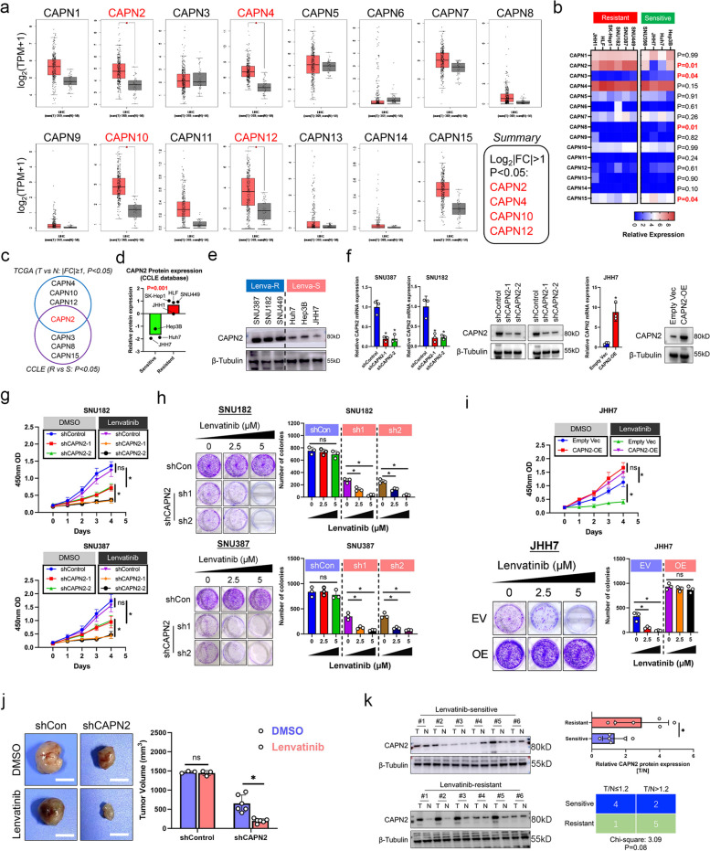
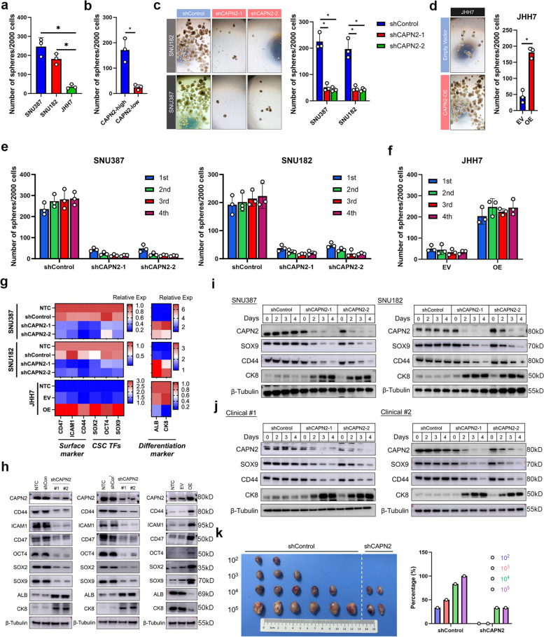
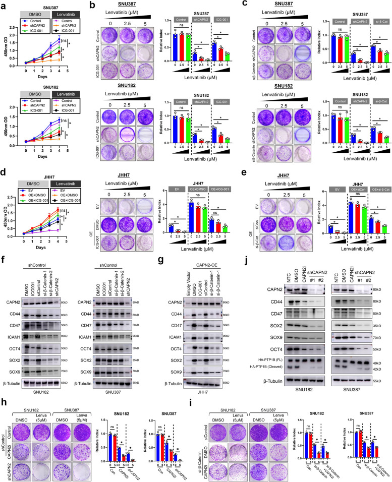
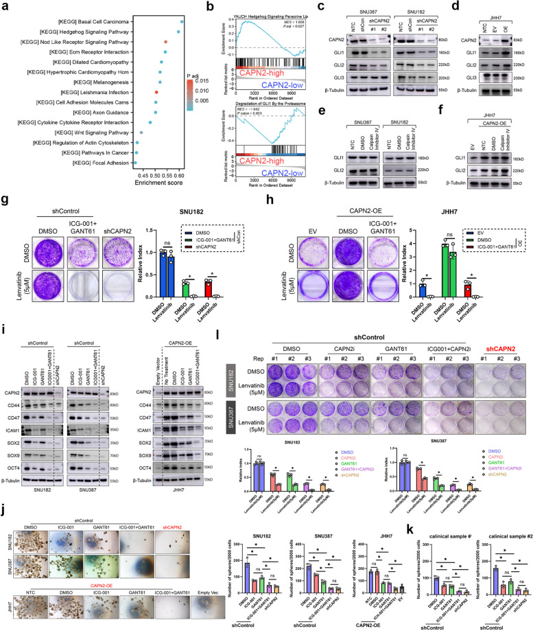
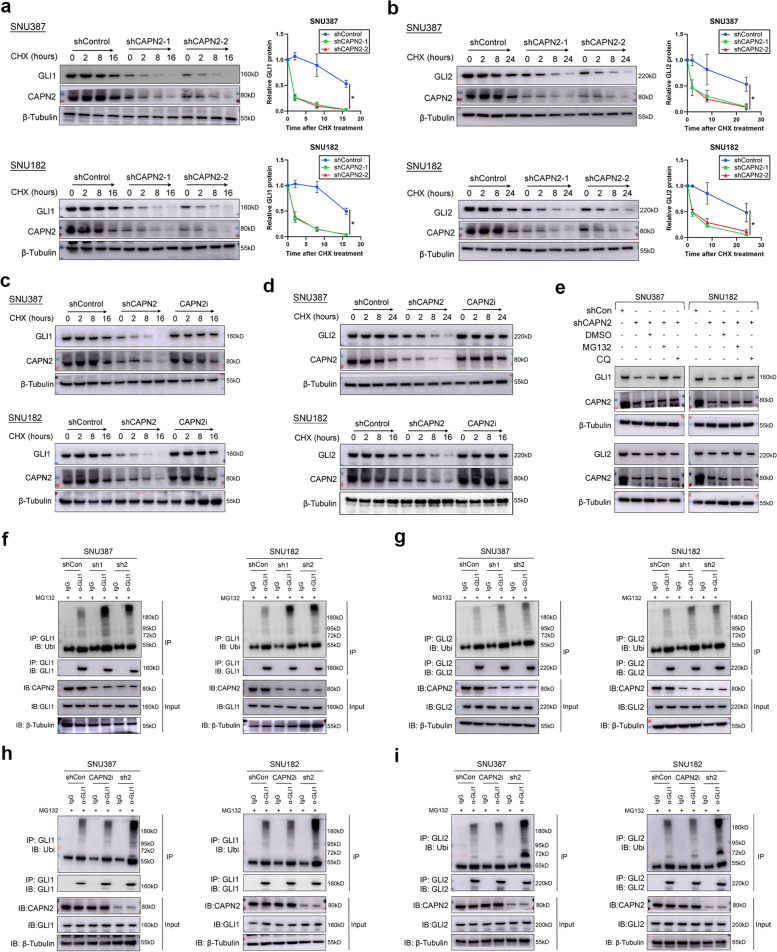
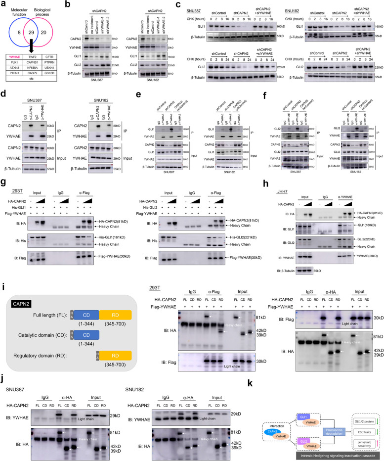
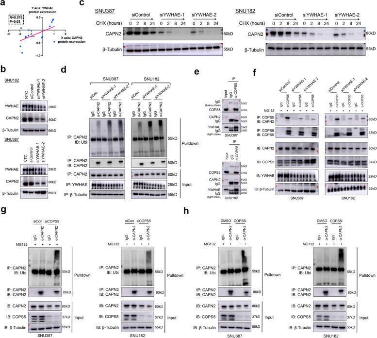
Lenvatinib, an approved first-line regimen, has been widely applied in hepatocellular carcinoma (HCC). However, clinical response towards Lenvatinib was limited, emphasizing the importance of understanding the underlying mechanism of its resistance. Herein, we employed integrated bioinformatic analysis to identify calpain-2 (CAPN2) as a novel key regulator for Lenvatinib resistance in HCC, and its expression greatly increased in both Lenvatinib-resistant HCC cell lines and clinical samples. Further in vitro and in vivo experiments indicated that knocking down CAPN2 greatly sensitized HCC cells to Lenvatinib treatment, while overexpression of CAPN2 achieved opposite effects in a Lenvatinib-sensitive HCC cell line. Interestingly, we observed a close relationship between CAPN2 expression and cancer stem cell (CSC) traits in HCC cells, evidenced by impaired sphere-forming and CSC-related marker expressions after CAPN2 knockdown, and verse vice. Mechanistically, we strikingly discovered that CAPN2 exerted its function by both enzyme-dependent and enzyme-independent manner simultaneously: activating β-Catenin signaling through its enzyme activity, and preventing GLI1/GLI2 degradation through direct binding to YWHAE in an enzyme-independent manner, which disrupting the association between YWHAE and GLI1/GLI2 to inhibit YWHAE-induced degradation of GLIs. Notably, further co-immunoprecipitation assays revealed that YWHAE could promote the protein stability of CAPN2 via recruiting a deubiquitinase COPS5 to prevent ubiquitination-induced degradation of CAPN2. In summary, our data demonstrated that CAPN2 promoted Lenvatinib resistance via both catalytic activity-dependent and -independent approaches. Reducing CAPN2 protein rather than inhibiting its activity might be a promising strategy to improve Lenvatinib treatment efficiency in HCC.


Conflict of interest statement

Declarations. Ethics approval and consent to participate: This study was approved by the Fudan University Shanghai Cancer Center Research Ethics Committee (Approved Number: 050432–4-2108*), and all individuals provided informed consent for inclusion of their tissue in this study. Moreover, animal study was approved by the Animal Experimentation Ethics Committee of Shanghai Cancer Center, Fudan University (Approved Number: FUSCC-IACUC-S2022-0209). Competing interests: The authors declare that they have no competing interests.

Figures

Fig. 1
Fig. 1
Identification of CAPN2 as the key member of calpain family to promote Lenvatinib resistance in HCC. a Expression patterns of each calpain family member between HCC and normal liver tissues according to TCGA dataset; Data were collected from GEPIA database. b Expression patterns of each calpain family member between Lenvatinib-resistant and Lenvatinib-sensitive HCC cell lines according to CCLE dataset. c Overlapped genes between the screening results of TCGA and CCLE dataset mentioned in (a) and (b). d Comparison of CAPN2 protein expression between indicated HCC cell lines according to CCLE dataset. e WB assay for detecting CAPN2 expression in indicated HCC cell lines. f PCR and immunoblotting assay results for verifying the CAPN2 knockdown efficiencies in indicated HCC cell lines. g CCK8 assay results for determining the effects of CAPN2 knockdown on Lenvatinib response in indicated HCC cell lines. h Colony formation assay results for determining the effects of CAPN2 knockdown on Lenvatinib response in indicated HCC cell lines. i CCK8 (upper) and colony formation (lower) assay results for determining the effects of CAPN2 overexpression on Lenvatinib response in indicated HCC cell lines. j In vivo results of CAPN2 knockdown on Lenvatinib response in SNU387 cells; for shControl group, 3 mice per group; for shCAPN2 group, 6 mice per group; Initially, 5 × 106 cells per mouse were injected subcutaneously into the right posterior flanks of 6-week-old BALB/c nude mice; After tumor establishment, mice were randomly assigned to 5 days per week treatment with vehicle, Lenvatinib (4 mg kg − 1, oral gavage). k Immunoblotting assays results of CAPN2 protein expression in Lenvatinib-resistant and Lenvatinib-sensitive clinical samples. “ns” indicates no significance; “*” indicates P value less than 0.05
Fig. 2
Fig. 2
CAPN2 induced CSC traits in HCC. a Sphere-forming capacities of indicated HCC cell lines according to sphere-forming experiments. b Sphere-forming capacities of indicated cells derived from clinical samples according to sphere-forming experiments (CAPN2-high: 3 samples; CAPN2-low 3 samples). c Effects of CAPN2 knockdown on sphere-forming capacities of indicated HCC cell lines. d Effects of CAPN2 overexpression on sphere-forming capacities of JHH7 cells. e Effects of CAPN2 knockdown on serial sphere-forming capacities of indicated HCC cell lines. f Effects of CAPN2 overexpression on serial sphere-forming capacities of indicated HCC cell lines. g PCR assay results for the changes of indicated CSC-related marker and liver differentiation marker expressions upon CAPN2 expression alterations; experiments were conducted in triplicate. h WB assay results for the dynamic changes of indicated CSC-related marker and liver differentiation marker expressions upon CAPN2 knockdown (left two panel) or overexpression (right panel). i WB assay results for the dynamic changes of CSC-related marker and liver differentiation marker expressions after receiving shRNAs targeting CAPN2 in indicated cell lines. j WB assay results for the dynamic changes of CSC-related marker and liver differentiation marker expressions after receiving shRNAs targeting CAPN2 in sphere cells derived from indicated clinical samples. k Limiting dilution xenograft assay results of SNU387 cells received indicated treatment; Initially, indicated number of SNU387 cells per mouse were injected subcutaneously into the right posterior flanks of 6-week-old BALB/c nude mice (n = 6 per group), tumors were harvested 6 weeks after initial transplantation, and the frequence were calculated. “ns” indicates no significance; “*” indicates P value less than 0.05
Fig. 3
Fig. 3
CAPN2 partially relied on β-Catenin signaling in an enzyme-dependent manner. a CCK8 assay results for determining the effects of β-Catenin signaling inhibition on Lenvatinib response of indicated HCC cell lines; CAPN2 knockdown was used as positive controls. b Colony formation assay results for determining the effects of β-Catenin signaling inhibition on Lenvatinib response of indicated HCC cell lines; CAPN2 knockdown was used as positive controls. c Colony formation assay results for determining the effects of silencing β-Catenin expression on Lenvatinib response of indicated HCC cell lines; CAPN2 knockdown was used as positive controls. d CCK8 (left) and colony formation (right) assay results for determining the effects of β-Catenin signaling inhibition on Lenvatinib response in CAPN2-overexpressed JHH7 cells. e Colony formation assay results for determining the effects of silencing β-Catenin expression on Lenvatinib response in CAPN2-overexpressed JHH7 cells. f WB assay results to evaluate the CSC-related expression alterations upon receiving indicated treatments in SNU182 (left) and SNU387 (right) cells. g WB assay results for evaluating CSC-related expression alterations upon receiving indicated treatments in JHH7 cells. h Comparison of inhibitory effects of SNU387 and SNU182 cells received indicated treatments; revealed weaker inhibitory effects of CAPN2 inhibitor than CAPN2 knockdown did. i Comparison of inhibitory effects of SNU387 and SNU182 cells received indicated treatments; revealed CAPN2 inhibitor exerted no further inhibitory effects when β-Catenin expression was silenced. j WB assay experiments for comparing the dynamic changes of CSC-related marker expression between CAPN2 knockdown and CAPN2 enzymatic inhibition. “ns” indicates no significance; “*” indicates P value less than 0.05
Fig. 4
Fig. 4
CAPN2 relied on Hedgehog signaling to promote Lenvatinib resistance in an enzyme-independent manner. a KEGG analysis revealed Hedgehog signaling as the most significant enriched pathway in CAPN2-high group in TCGA LIHC dataset. b Gene set enrichment analysis revealed Hedgehog signature was enriched in CAPN2-high group in TCGA LIHC dataset. c Effects of CAPN2 knockdown on the GLIs protein expressions according to WB assays. d Effects of CAPN2 overexpression on the GLIs protein expressions according to WB assays. e Effects of CAPN2 inhibitor on the GLIs protein expressions according to immunoblotting assays. f Effects of CAPN2 inhibitor on the GLIs protein expressions in CAPN2-overexpressed JHH7 cells according to WB assays. g Colony formation assays demonstrated the syngenetic effects of GANT61 and ICG-001 in sensitizing SNU387 cells to Lenvatinib. h Colony formation assays demonstrated the syngenetic effects of GANT61 and ICG-001 in abolishing the effects of CAPN2 overexpression on Lenvatinib in JHH7 cells. i WB experiment results confirmed the syngenetic effects of GANT61 and ICG-001 in decreasing CSC-related marker expressions in CAPN2-high (SNU182 and SNU387) and CAPN2-overexpressed JHH7 cells. j Sphere-forming assay results of HCC cell lines received indicated treatments; Representative images were shown in the left panel. k Sphere-forming assay results of HCC clinical samples received indicated treatments. l Colony formation assay for evaluating the potential syngenetic effects of GANT61 and CAPN2 inhibitor on reversing Lenvatinib resistance in CAPN2-high (SNU182 and SNU387) cells; Experiments were conducted in triplicate. “ns” indicates no significance; “*” indicates P value less than 0.05
Fig. 5
Fig. 5
CAPN2 promoted Hedgehog signaling via preventing GLI1/2 proteasome degradation. a Half-life alterations of GLI1 protein upon CAPN2 knockdown were determined by CHX-chasing experiments. b Half-life alterations of GLI2 protein upon CAPN2 knockdown were determined by CHX-chasing experiments. c Half-life alterations of GLI1 protein upon CAPN2 inhibitor treatment were determined by CHX-chasing experiments. d Half-life alterations of GLI2 protein upon CAPN2 inhibitor treatment were determined by CHX-chasing experiments. e Recovery effects of proteasome pathway inhibitor, MG132, and lysosome pathway inhibitor, chloroquine, on GLI1/2 protein expressions in CAPN2 knockdown HCC cells were determined by immunoblotting assays. f Ubiquitination experiments for detecting the ubiquitination levels of GLI1 in HCC cells received indicated treatments. g Ubiquitination experiments for detecting the ubiquitination levels of GLI2 in HCC cells received indicated treatments. h Ubiquitination experiments for detecting the changes of ubiquitination levels of GLI1 in HCC cells received CAPN2 inhibitor and specific shRNA. i Ubiquitination experiments for detecting the changes of ubiquitination levels of GLI2 in HCC cells received CAPN2 inhibitor and specific shRNA. “ns” indicates no significance; “*” indicates P value less than 0.05
Fig. 6
Fig. 6
CAPN2 binds to YWHAE to prevent YWHAE-GLI1/2 interaction and following GLI1/2 degradation. a Candidates of CAPN2 binding partners according to PINA 3.0 dataset; a typical negative GLI1, GLI2 regulator, YWHAE, was listed. b WB assay results of GLI1 and GLI2 protein upon silencing YWHAE in CAPN2-knockdown HCC cells. C Half-life span changes of GLI1 and GLI2 protein upon receiving indicated treatments in HCC cells were determined by CHX chasing experiments. d Co-IP assays confirmed the interaction between CAPN2 and YWHAE. e Co-IP assays revealed CAPN2 knockdown promote YWHAE-GLI1 interaction, while CAPN2 inhibitor shed no effects on this interaction in HCC cells. f Co-IP assays revealed CAPN2 knockdown promoted YWHAE-GLI2 interaction, while CAPN2 inhibitor shed no effects on this interaction in HCC cells. g Co-transfection of HA-tagged CAPN2, His-tagged Gli1 and Flag-tagged YWHAE in 293 T cells followed by co-IP using anti-Flag antibodies to determine the effects of CAPN2 on YWHAE-GLI1 and GLI2 interactions; Cells were pre-treated with MG132 for 5 h to avoid GLI1/2 degradation. h Exogenous expression of different levels of CAPN2 in JHH7 cells followed by co-IP experiments to determine the effects of CAPN2 on YWHAE-GLI1 and GLI2 interactions; Cells were pre-treated with MG132 for 5 h to avoid GLI1/2 degradation. i Schematic diagram of CAPN2 protein domains (left) and Co-IP experiments for evaluating the critical domain responsible for the interaction between CAPN2 and YWHAE. j Co-IP experiments for validating the critical domain responsible for the interaction between CAPN2 and YWHAE in HCC cells. k Schematic diagram of CAPN2-mediated Hedgehog signaling activation in enzyme-independent manner
Fig. 7
Fig. 7
YWHAE binding CAPN2 to promote its protein stability via recruiting COPS5. a Positive correlation between YWHAE and CAPN2 proteins according to CCLE dataset. b Effects of YWHAE knockdown on CAPN2 expression in SNU182 (upper) and SNU387 (lower) cells. c Half-life span of CAPN2 protein upon YWHAE knockdown in HCC cells was determined by CHX chasing experiments. d Ubiquitination experiments for detecting the ubiquitination levels of CAPN2 in HCC cells received indicated treatments. e Co-IP experiments indicated interaction among COPS5, CAPN2 and YWHAE in SNU182 and SNU387 cells; endogenous protein was immunoprecipitated via using anti-COPS5 antibody, followed by WB assays. f Co-IP experiments indicated silencing YWHAE abolished COPS5-CAPN2 interaction. g Ubiquitination experiments for detecting the ubiquitination levels of CAPN2 in HCC cells upon COPS5 knockdown. h Ubiquitination experiments for detecting the ubiquitination levels of CAPN2 in HCC cells upon COPS5 inhibition
Fig. 8
Fig. 8
Diagram illustration of present study. CAPN2 exerted its function through promoting β-Catenin and Hedgehog signaling simultaneously: 1) activating β-Catenin signaling through its enzyme activity, and 2) preventing GLI1/GLI2 degradation through direct binding to YWHAE in an enzyme-independent manner, which disrupts the association between YWHAE and GLI1/GLI2 to inhibit YWHAE-induced degradation of GLIs. Moreover, intracellular CAPN2 protein stability was also enhanced by YWHAE binding, as it could serve as a scaffold to recruit deubiquitinase COPS5 to prevent proteasome degradation of CAPN2

Similar articles

References

    1. Lehrich BM, Zhang J, Monga SP, Dhanasekaran R. Battle of the biopsies: Role of tissue and liquid biopsy in hepatocellular carcinoma. J Hepatol. 2024;80(3):515–30. 10.1016/j.jhep.2023.11.030. - PMC - PubMed
    1. Han X, Sun Q, Xu M, Zhu GH, Gao RK, Ni BY, et al. Semin liver. Dis unraveling the complexities of immune checkpoint inhibitors in hepatocellular carcinoma. Semin Liver Dis. 2023;43(4):383–401. 10.1055/s-0043-1776127. - PubMed
    1. Simon SM. Fighting rare cancers: lessons from fibrolamellar hepatocellular carcinoma. Nat Rev Cancer. 2023;23(5):335–46. 10.1038/s41568-023-00554-w. - PMC - PubMed
    1. Jiang J, Huang H, Chen R, Lin Y, Ling Q. Immunotherapy for hepatocellular carcinoma recurrence after liver transplantation, can we harness the power of immune checkpoint inhibitors? Front Immunol. 2023;14:1092401. 10.3389/fimmu.2023.1092401. - PMC - PubMed
    1. Su TH, Wu CH, Liu TH, Ho CM, Liu CJ. Clinical practice guidelines and real-life practice in hepatocellular carcinoma: a Taiwan perspective. Clin Mol Hepatol. 2023;29(2):230–41. 10.3350/cmh.2022.0421. - PMC - PubMed

Publication types

MeSH terms

LinkOut - more resources