

Sustained Vascular Inflammatory Effects of SARS-CoV-2 Spike Protein on Human Endothelial Cells
- PMID: 39739157
- PMCID: PMC12336097
- DOI: 10.1007/s10753-024-02208-x
Sustained Vascular Inflammatory Effects of SARS-CoV-2 Spike Protein on Human Endothelial Cells
Abstract
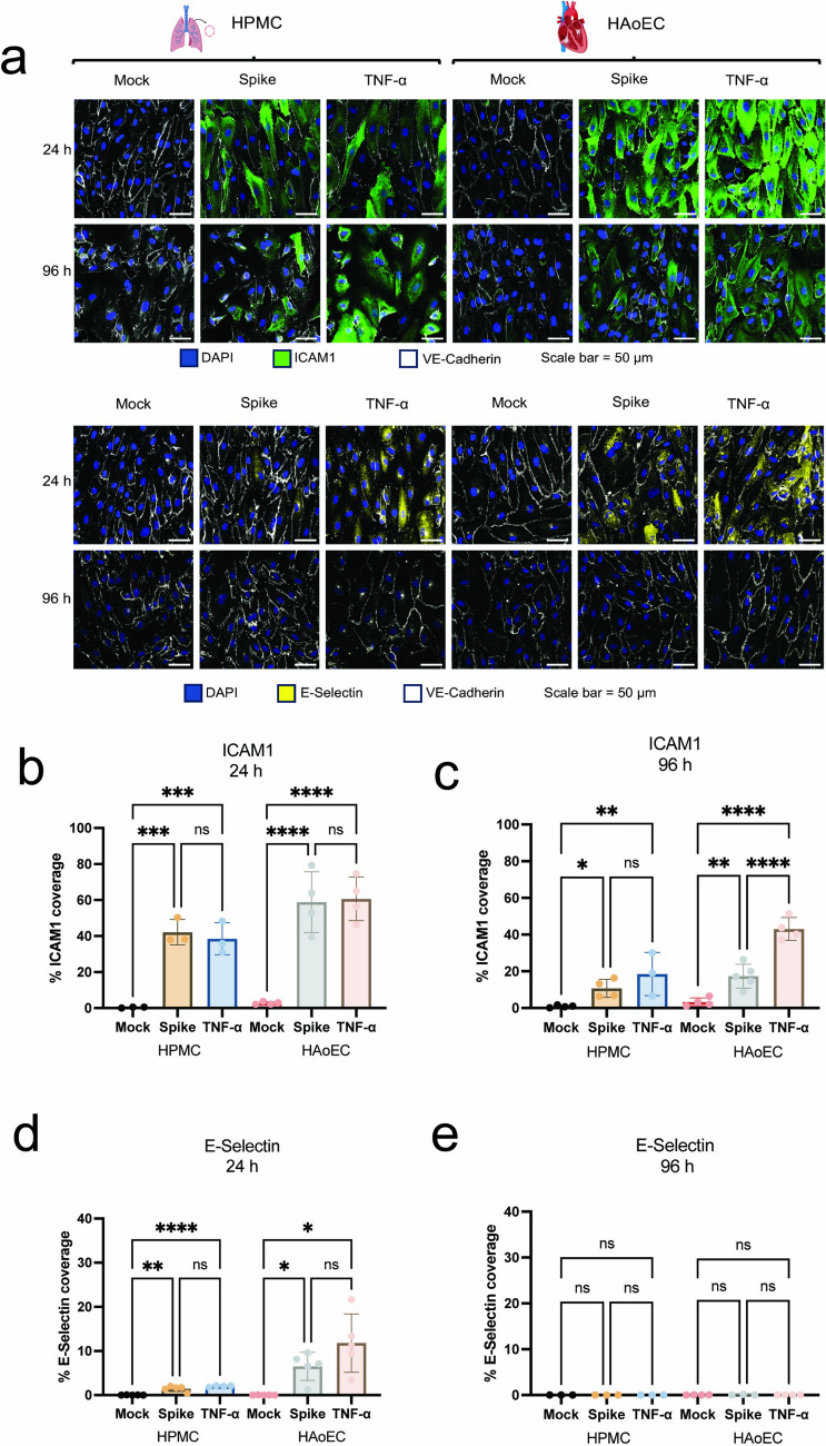
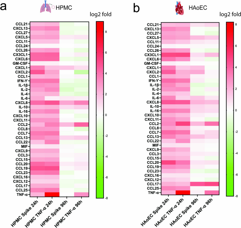
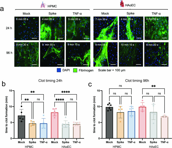
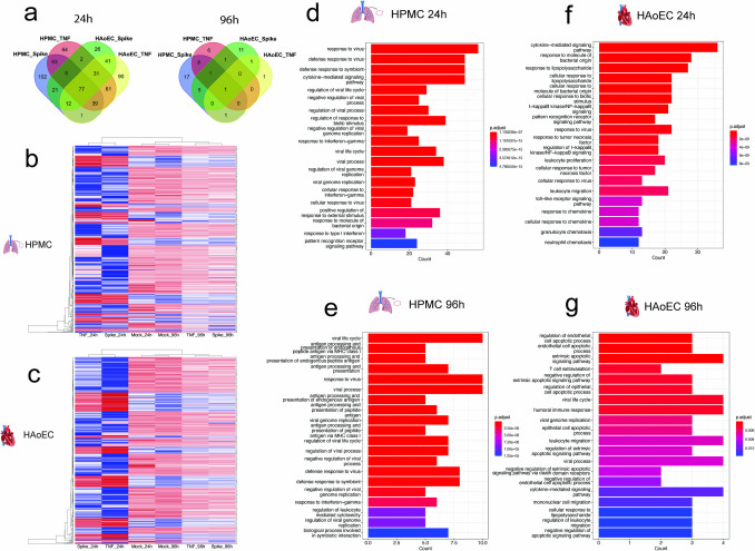
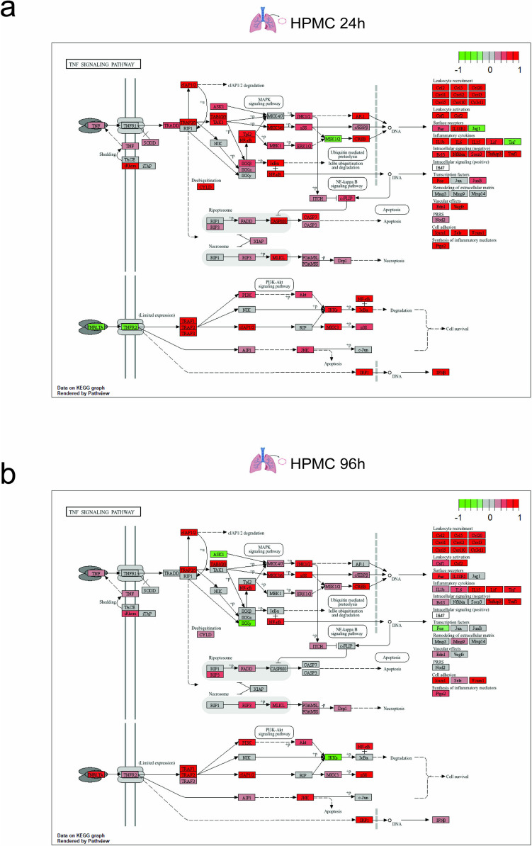
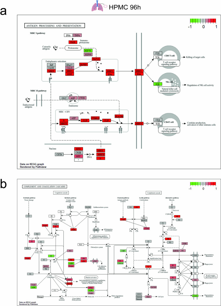
Severe acute respiratory syndrome coronavirus 2 (SARS-CoV-2) infection has been associated with systemic inflammation and vascular injury, which contribute to the development of acute respiratory syndrome (ARDS) and the mortality of COVID-19 infection. Moreover, multiorgan complications due to persistent endothelial dysfunction have been suspected as the cause of post-acute sequelae of SARS-CoV-2 infection. Therefore, elucidation of the vascular inflammatory effect of SARS-CoV-2 will increase our understanding of how endothelial cells (ECs) contribute to the short- and long-term consequences of SARS-CoV-2 infection. Here, we investigated the interaction of SARS-CoV-2 spike protein with human ECs from aortic (HAoEC) and pulmonary microvascular (HPMC) origins, cultured under physiological flow conditions. We showed that the SARS-CoV-2 spike protein triggers prolonged expression of cell adhesion markers in both ECs, similar to the effect of TNF-α. SARS-CoV-2 spike treatment also led to the release of various cytokines and chemokines observed in severe COVID-19 patients. Moreover, increased binding of leucocytes to the endothelial surface and a procoagulant state of the endothelium were observed. Transcriptomic profiles of SARS-CoV-2 spike-activated HPMC and HAoEC showed prolonged upregulation of genes and pathways associated with responses to virus, cytokine-mediated signaling, pattern recognition, as well as complement and coagulation pathways. Our findings support experimental and clinical observations of the vascular consequences of SARS-CoV-2 infection and highlight the importance of EC protection as one of the strategies to mitigate the severe effects as well as the possible post-acute complications of COVID-19 disease.
Keywords: Endothelial cells; SARS-CoV-2; Spike protein; Vascular inflammation.
© 2024. The Author(s).
Conflict of interest statement
Declarations. Competing Interests: The authors declare no competing interests.
Figures

Similar articles
-
The fibrin-derived peptide FX06 protects human pulmonary endothelial cells against the COVID-19-triggered cytokine storm.Front Immunol. 2025 Jun 19;16:1591860. doi: 10.3389/fimmu.2025.1591860. eCollection 2025. Front Immunol. 2025. PMID: 40612940 Free PMC article.
-
Site specific N- and O-glycosylation mapping of the spike proteins of SARS-CoV-2 variants of concern.Sci Rep. 2023 Jun 21;13(1):10053. doi: 10.1038/s41598-023-33088-0. Sci Rep. 2023. PMID: 37344512 Free PMC article.
-
Determinants of susceptibility to SARS-CoV-2 infection in murine ACE2.J Virol. 2025 Jun 17;99(6):e0054325. doi: 10.1128/jvi.00543-25. Epub 2025 May 12. J Virol. 2025. PMID: 40353671 Free PMC article.
-
Measures implemented in the school setting to contain the COVID-19 pandemic.Cochrane Database Syst Rev. 2022 Jan 17;1(1):CD015029. doi: 10.1002/14651858.CD015029. Cochrane Database Syst Rev. 2022. Update in: Cochrane Database Syst Rev. 2024 May 2;5:CD015029. doi: 10.1002/14651858.CD015029.pub2. PMID: 35037252 Free PMC article. Updated.
-
Antibody tests for identification of current and past infection with SARS-CoV-2.Cochrane Database Syst Rev. 2022 Nov 17;11(11):CD013652. doi: 10.1002/14651858.CD013652.pub2. Cochrane Database Syst Rev. 2022. PMID: 36394900 Free PMC article.
Cited by
-
Long COVID Mechanisms, Microvascular Effects, and Evaluation Based on Incidence.Life (Basel). 2025 May 30;15(6):887. doi: 10.3390/life15060887. Life (Basel). 2025. PMID: 40566540 Free PMC article. Review.
-
Integrated bioinformatics and experimental analysis of mitochondrial-associated membrane function and mechanism in acute respiratory distress syndrome.Sci Rep. 2025 Jul 9;15(1):24602. doi: 10.1038/s41598-025-10405-3. Sci Rep. 2025. PMID: 40634559 Free PMC article.
References
MeSH terms
Substances
Grants and funding
LinkOut - more resources
Full Text Sources
Medical
Miscellaneous
