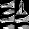
Retrospective evaluation of computed tomographic-guided Tru-Cut biopsies in 16 dogs and 14 cats with nasal cavity mass lesions
- PMID: 39739338
- PMCID: PMC11683400
- DOI: 10.1111/jvim.17296
Retrospective evaluation of computed tomographic-guided Tru-Cut biopsies in 16 dogs and 14 cats with nasal cavity mass lesions
Abstract
Background: Approximately 80% of nasal masses in dogs and 91% of nasal masses in cats are reported to be malignant, but the currently reported diagnostic rate of neoplasia is 54% using blind or rhinoscopic biopsy techniques.
Hypothesis/objectives: Describe the technique of computed tomography (CT)-guided Tru-Cut (Tru-Cut biopsy needle, Merit Medical Systems, Utah, USA) nasal biopsies in cats and dogs to determine the diagnostic rate of neoplasia on the first round of sampling and to evaluate the safety of the technique.
Animals: Thirty client-owned animals, 16 dogs and 14 cats, that had CT-guided nasal biopsies performed to investigate nasal masses.
Methods: Retrospective, single-center, medical record review of 16 dogs and 14 cats that had CT-guided nasal biopsies performed between 2022 and 2024.
Results: Diagnostic biopsy samples were acquired using CT-guided Tru-Cut sampling in 28/30 cases (93%). The diagnosis was considered clinically appropriate in 26/30 cases (87%): neoplasia in 24/30 cases (80%) and rhinitis in 2/30 cases (7%). Neoplasia was the final diagnosis in 14/16 dogs (88%) and 10/14 cats (71%).
Conclusions and clinical importance: Computed tomographic-guided Tru-Cut biopsies can result in a high first-round diagnosis of neoplasia in nasal masses in cats and dogs, without clinically relevant complications. This technique is a useful alternative method of sampling nasal masses that may be difficult to access via rhinoscopy.
Keywords: biopsy; canine; feline; histology; nasopharynx; neoplasia; rhinoscopy.
© 2024 The Author(s). Journal of Veterinary Internal Medicine published by Wiley Periodicals LLC on behalf of American College of Veterinary Internal Medicine.
Conflict of interest statement
All authors are currently employed as veterinarians by Linnaeus Veterinary Limited. Linnaeus Veterinary Limited did not have any influence on the study. All histopathology was performed by an external laboratory, therefore no bias associated with this study.
Figures


References
-
- Madewell BR, Priester WA, Gillette EL, Snyder SP. Neoplasms of the nasal passages and paranasal sinuses in domesticated animals as reported by 13 veterinary colleges. Am J Vet Res. 1976;37(7):851‐856. - PubMed
-
- Lana S, Turek M. Nasal cavity and sinus tumors. In: Vail D, Thamm D, Liptak J, eds. Withrow and MacEwen's Small Animal Clinical Oncology. 6th ed. St. Louis, MO: Elsevier; 2019:494‐504.
-
- Harris BJ, Lourenço BN, Dobson JM, Herrtage ME. Diagnostic accuracy of three biopsy techniques in 117 dogs with intra‐nasal neoplasia. J Small Anim Pract. 2014;55(4):219‐224. - PubMed
-
- Miles MS, Dhaliwal RS, Moore MP, Reed AL. Association of magnetic resonance imaging findings and histologic diagnosis in dogs with nasal disease: 78 cases (2001‐2004). J Am Vet Med Assoc. 2008;232(12):1844‐1849. - PubMed
MeSH terms
Grants and funding
LinkOut - more resources
Full Text Sources
Medical
Miscellaneous

