Review
. 2025 Mar 1;328(3):H622-H638.
doi: 10.1152/ajpheart.00277.2024. Epub 2024 Dec 31.

The role of mechanosignaling in the control of myocardial mass

Affiliations
Review

The role of mechanosignaling in the control of myocardial mass

Maicon Landim-Vieira et al. Am J Physiol Heart Circ Physiol. .

Abstract

Regulation of myocardial mass is key for maintaining cardiovascular health. This review highlights the complex and regulatory relationship between mechanosignaling and myocardial mass, influenced by many internal and external factors including hemodynamic and microgravity, respectively. The heart is a dynamic organ constantly adapting to changes in workload (preload and afterload) and mechanical stress exerted on the myocardium, influencing both physiological adaptations and pathological remodeling. Mechanosignaling pathways, such as the mitogen-activated protein kinases (MAPKs) and the phosphoinositide 3-kinases and serine/threonine kinase (PI3K/Akt) pathways, mediate downstream effects on gene expression and play key roles in transducing mechanical cues into biochemical signals, thereby modulating cellular processes, including control of myocardial mass. Dysregulation of these processes can lead to pathological cardiac remodeling, such as hypertrophic cardiomyopathy. Furthermore, recent studies have highlighted the importance of protein quality control mechanisms, such as the ubiquitin-proteasome system, in settings of extreme physiological conditions that alter the heart workload such as pregnancy and microgravity. Overall, this review provides a thorough insight into how mechanical signals are converted into chemical signals to regulate myocardial mass in both healthy and diseased conditions.

Keywords: mechanotransduction; misfolding; proteasome; protein degradation; protein homeostasis.


Conflict of interest statement

DISCLOSURES

No conflicts of interest, financial or otherwise, are declared by the authors.

Figures

Figure 1.
Figure 1.. Intracellular signals triggered by mechanical loads at the costameres.
Mechanical deformation of the cellular substrate is internalized by increased tension via integrin anchorage to the sarcolemma that elicits activation of mitogen-related protein kinase (MAPK) signaling. MAPK signaling may be initiated by recruitment and activation of costameric proteins including talin, vinculin, paxillin, FAK, and melusin. FAK phosphorylates targets Raf to activate MEK and ERK1/2. Alternatively, FAK phosphorylates p85a to activate the PI3K-AKT-mTORC1-S6K signaling cascade. Continuous arrows indicate mechanical stimulation while dashed arrows indicate biochemical signals.
Figure 2.
Figure 2.. Angiotensin type 1 receptor is a channel with dual extracellular signal transduction via ligand and mechanical stress.
The canonical activation of the angiotensin type 1 receptor (AGTR1) by angiotensin II leads to ERK1/2 activation via Gαq and MAPK as well as PKC and phosphoinositide signaling via PLC-β4. Mechanical activation of the angiotensin type 1 receptor is distinct from the canonical angiotensin II agonist signaling in recruiting Gαi subunit recruitment and mitogen signaling activation.
Figure 3.
Figure 3.. Mechanically activated ion channels drive ion influx.
Mechanically activated channels lying at the cellular membrane like PIEZO (PIEZO 1, PIEZO2) and L-type ion channels drive ion influx such as Ca2+, as well as Na+ in the case of PIEZO, to increase functions like smooth muscle contraction, endothelial signaling, and Ca2+-induced Ca2+ release.
Figure 4.
Figure 4.. Load-driven signals at the myofilaments.
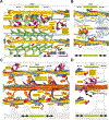
(A) Low Z-disc tension (top) favors Z-disc disassembly and ubiquitination of sarcomeric proteins via upstream upregulation of ubiquitin ligases like MuRF1/2. High Z-disc tension (bottom) drives myofilament assembly via increased CapZ dynamics and release of MLP and NFAT nuclear migration via calcineurin dephosphorylation. (B) The I-band under high mechanical tension (bottom) recruits an ERK/12 signaling cascade coordinated by FHL1 interacting with the titin PEVK domain while under low mechanical tension this signaling is not activated (top). (C) Under low mechanical tension the A-band proteins are more susceptible to ubiquitination (top) while under high mechanical tension sarcomeric proteins may become more phosphorylated in response to upstream activation of kinases. (D) Titin ubiquitination at the M-line facilitated by MuRF1/2 recruitment to a site proximal to the titin kinase domain (top) but under high mechanical tension MuRF1/2 is more distant to its target ubiquitination sites due to unfolding of a spacer domain between the MuRF1/2 binding site and the titin kinase domain.
Figure 5.
Figure 5.. Mechanical feedback redistributes the balance between protein synthesis and degradation.
Atrophic signaling increases ubiquitination of proteins and subsequent degradation by the proteasome as well as translocation of the transcription factor FoxO1/3 to the nucleus (left). Increased mechanical feedback (hypertrophic signaling) activates ribosome translation via S6K1 and RSK3 phosphorylation of the ribosomal S6 protein and increased phosphorylation of FoxO1/3 for retention in the cytoplasm (right).
Figure 6.
Figure 6.. Increased mechanical signaling is associated with pro-hypertrophic genes while atrophy signaling activates a distinctive subset of genes.
Transcriptional activation of SRF via RSK3, NFκ-B and GATA via ERK1/2, and NFAT via calcineurin signaling in response to increased mechanical feedback while FoxO1/3 and SMAD promote expression of atrogenes with muscle disuse.

Similar articles

References

    1. Lyon RC, Zanella F, Omens JH, andSheikh F (2015) Mechanotransduction in cardiac hypertrophy and failure Circ Res 116, 1462–1476 10.1161/CIRCRESAHA.116.304937 - DOI - PMC - PubMed
    1. Cui M, Wang Z, Bassel-Duby R, andOlson EN (2018) Genetic and epigenetic regulation of cardiomyocytes in development, regeneration and disease Development 145, 10.1242/dev.171983 - DOI - PMC - PubMed
    1. Vigil-Garcia M, Demkes CJ, Eding JEC, Versteeg D, de Ruiter H, Perini I et al. (2021) Gene expression profiling of hypertrophic cardiomyocytes identifies new players in pathological remodelling Cardiovasc Res 117, 1532–1545 10.1093/cvr/cvaa233 - DOI - PMC - PubMed
    1. Gordan R, Gwathmey JK, andXie LH (2015) Autonomic and endocrine control of cardiovascular function World J Cardiol 7, 204–214 10.4330/wjc.v7.i4.204 - DOI - PMC - PubMed
    1. Tirziu D, Giordano FJ, andSimons M (2010) Cell communications in the heart Circulation 122, 928–937 10.1161/CIRCULATIONAHA.108.847731 - DOI - PMC - PubMed

LinkOut - more resources