. 2025 Aug;12(29):e2406510.
doi: 10.1002/advs.202406510. Epub 2024 Dec 30.

Viral-Mediated Connexin 26 Expression Combined with Dexamethasone Rescues Hearing in a Conditional Gjb2 Null Mice Model

Affiliations

Viral-Mediated Connexin 26 Expression Combined with Dexamethasone Rescues Hearing in a Conditional Gjb2 Null Mice Model

Xiaohui Wang et al. Adv Sci (Weinh). 2025 Aug.

Abstract

GJB2 encodes connexin 26 (Cx26), the most commonly mutated gene causing hereditary non-syndromic hearing loss. Cx26 is mainly expressed in supporting cells (SCs) and fibrocytes in the mammalian cochlea. Gene therapy is currently considered the most promising strategy for eradicating genetic diseases. However, there have been no significant effects of gene therapy for GJB2 gene mutation-associated deafness because deficiency of Cx26 leads to expanded sensory epithelial damage. In this study, the AAV2.7m8 serotype combined with the gfaABC1D promoter targeted infection of SCs is identified. It is found that Gjb2 gene replacement therapy in wild-type mice results in sensory hair cells (HCs) deficits, excessive inflammatory responses, and hearing loss. This may be one of the key factors contributing to the hardship of GJB2 gene replacement therapy. Dexamethasone (DEX) shows promising results in inhibiting macrophage recruitment, with a protective effect against HC damage. Further, the combination of AAV2.7m8-Gjb2 with DEX shows a synergistic effect and enhances the gene therapy effect in a conditional Cx26 null mice model. These results indicate that the combination of gene therapy and medication will provide a new strategy for the treatment of hereditary deafness associated with GJB2 defects.

Keywords: AAV2.7m8; GJB2; dexamethasone; gene therapy; hearing loss.


Conflict of interest statement

The authors declare no conflict of interest.

Figures

Figure 1
Figure 1
AAV‐2.7m8‐ gfaABC1D‐eGFP efficiently transduced cochlear SCs. A) Schematics of the AAV‐2.7m8‐ gfaABC1D‐eGFP genome organization. B) Process diagram of AAVs in vivo and in vitro infection of the inner ear. AAVs serotypes at the same dose (5 × 10⁹ genome‐containing (particles) (GCs) per ear). Cochleae were harvested at P21 after microinjection with 1.5 µL of AAV stock solution in one ear at P2. C,D) Representative images of eGFP fluorescence (green), Phalloidin (red), DAPI (blue), and Sox2(white) staining in the middle turns of cochleae infected with different AAVs serotypes in organotypic cochlear explants (C) and in vivo (D), respectively. Scale bar, 30 µm. (E‐H) Percentage of eGFP‐positive HCs and SCs per 100 µm corresponding to C‐D. Data are shown as mean ± SEM. Significance tests were performed between AAV‐2.7m8 and other AAV serotypes. * p < 0.05, ** p < 0.01, and *** p < 0.001. ns: non‐significant. N = 3–5 in each group. IHCs, inner hair cells; OHCs, outer hair cells; SCs, supporting cells.
Figure 2
Figure 2
AAV2.7m8‐gfaABC1D‐eGFP efficiently transduces different types of SCs in vivo. A) Representative images of supporting cells in apical, middle, and basal turns from the AAV2.7m8‐gfaABC1D‐eGFP (GFP, green) injected at a dose of 5 × 10⁹ genome‐containing (particles) (GCs) per ear of the mice at P2. White: Sox2; Green: GFP. Blue: DAPI. Scale bar, 30 µm. B–E) Infection efficiencies of AAV2.7m8‐gfaABC1D‐eGFP in DCs, OPCs, IPCs, and IPhC, respectively. Data are shown as mean ± SEM. N = 3 in each group. DCs, Deiters cells; OPCs, outer pillar cells; IPCs: outer pillar cells; IPhC: inner phalanges cells.
Figure 3
Figure 3
AAV2.7m8‐gfaABC1D‐eGFP is safe for the mouse inner ear. A) Diagram showing the administration mode of the AAV2.7m8‐gfaABC1D‐eGFP injection. WT mice were injected AAV2.7m8‐gfaABC1D‐eGFP at a dose of 5.0 × 10⁹ genome‐containing (particles) (GCs) per ear through RWM at P2. The mice were tested for ABR and sacrificed at P30. B) ABR waveforms were recorded in uninjected and AAV2.7m8‐eGFP injected mice. The ABR was evoked by 24 kHz tone bursts. C) Hearing thresholds of the uninjected and AAV2.7m8‐eGFP injected group at different frequencies. Data are shown as mean ± SEM. ns: non‐significant. N = 4 in each group. D,E) Statistics on the amplitude and latency of ABR wave I from (B) in uninjected and AAV2.7m8‐eGFP injected mice. Data are shown as mean ± SEM. ns: non‐significant. N = 4 in each group. F) Representative images of hair cells in apical, middle, and basal turns from the uninjected and AAV2.7m8‐eGFP injected mice at P30. Green, GFP fluorescence; Red: Phalliodin, Blue: DAPI. Scale bars = 30 µm. G) Quantification of HC loss at specific cochlear locations in the uninjected and AAV2.7m8‐eGFP injected groups. Mice injected with AAV2.7m8‐eGFP did not induce hair cell death compared to the uninjected group. Data are shown as mean ± SEM. N = 4 in each group.
Figure 4
Figure 4
AAV2.7m8‐Gjb2 successfully expresses Cx26 and the injection of AAV2.7m8‐Gjb2 into wild‐type (WT) mice leads to HC death and hearing loss, a process that can be inhibited by DEX. A) Schematic overview of the experimental process of AAV2.7m8‐Gjb2 injection and DEX administration of the WT mouse model. The mice were injected with AAV2.7m8‐gfaABC1D‐Gjb2 (5 × 10⁹ genome‐containing (particles) (GCs) per ear) at P2. Dexamethasone was administered to mice at a dose of 3 mg kg−1 every two days starting at P10‐24. All of the mice were tested for auditory brainstem response (ABR) and sacrificed at P24–P25. B) Cx26 (red) and GFP (green) immunolabeling in the middle turn of the WT and WT+AAV‐Gjb2 at a dose of 5 × 10⁹ genome‐containing (particles) (GCs) per ear of the mice at P2 respectively. White: phalloidin. Scale bars = 30 µm. C) Relative expression of Cx26 in different groups of SCs from B. Cx26‐positive cell expression rate has been used as a reference for WT mice. Data are shown as mean ± SEM. *** p < 0.001. N = 7 in each group. D) ABR waveforms were recorded in uninjected, AAV2.7m8‐Gjb2 and AAV2.7m8‐Gjb2+DEX administration mice. The mice were injected with vehicle or dexamethasone at a dose of 3 mg kg−1 every two days starting at P10‐24. The ABR was evoked by 24 kHz tone bursts. E) ABR thresholds of the uninjected, AAV2.7m8‐Gjb2 and AAV2.7m8‐Gjb2+DEX groups at different frequencies. Data are shown as mean ± SEM. * p < 0.05, ** p < 0.01, and *** p < 0.001. N = 5 in each group. F) ABR threshold shift in AAV2.7m8‐Gjb2 and AAV2.7m8‐Gjb2+DEX administration group at different frequencies. Data are shown as mean ± SEM. * p < 0.05, ** p < 0.01, and *** p < 0.001. ns: non‐significant. N = 5 in each group. G,H) Representative images and quantification of OHC loss at specific cochlear locations in the uninjected, AAV2.7m8‐Gjb2 and AAV2.7m8‐Gjb2+DEX groups. Scale bars = 50 µm. Data are shown as mean ± SEM. * p < 0.05, ** p < 0.01, and *** p < 0.001. ns: non‐significant. N = 5 in each group.
Figure 5
Figure 5
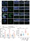
Distribution of CD45+ macrophages in AAV2.7m8‐Gjb2 injection group. Mice were injected with AAV2.7m8‐gfaABC1D‐eGFP or AAV2.7m8‐gfaABC1D‐Gjb2 (5 × 10⁹ genome‐containing (particles) (GCs) at P2 and the cochlea was harvested at P24. Beginning at P10‐24, mice were administered dexamethasone at a dose of 3 mg kg−1 once every two days. A–D) Immunofluorescent staining of CD45+ macrophages (green) in the apical, middle, and basal turns of different groups. Blue: DAPI. White: F‐actin. Scale bars represent 200 and 30 µm, respectively. E,F) Comparison of the numbers and the macrophage size of CD45+ cells in the different groups. Data are shown as mean ± SEM. * p < 0.05, ** p < 0.01, and *** p < 0.001. ns: non‐significant.
Figure 6
Figure 6
Rescue effect of the combination of AAV2.7m8‐Gjb2 and DEX in a conditional Gjb2 null mouse model. A) Schematic representation of the crossbreeding strategy to generate the targeted Gjb2‐null mouse model Fgfr3‐creER; Gjb2 loxp/loxp. B) Schematic overview of the experimental process of AAV2.7m8‐Gjb2 injection and DEX administration of the Fgfr3‐creER; Gjb2 loxp/loxp mouse model. The mice were injected subcutaneously with tamoxifen (TMX) at P0 and P1 and injected with AAV2.7m8‐gfaABC1D‐Gjb2 (5 × 10⁹ genome‐containing (particles) (GCs) per ear) at P2. Dexamethasone was administered to mice at a dose of 3 mg kg−1 every two days starting at P10‐24. All of the mice were tested for auditory brainstem response (ABR) and sacrificed at P24–P25. C) Cx26 (red) and GFP (green) immunolabeling in the middle turn of the Cx26‐null andCx26‐null+AAV‐Gjb2 at a dose of 5 × 10⁹ genome‐containing (particles) (GCs) per ear of the mice at P2 respectively. White: phalloidin. Scale bars = 30 µm. D) Relative expression of Cx26 in different groups of the SCs from C. Cx26‐positive cell expression rate has been used as a reference for WT mice. Data are shown as mean ± SEM. *** p < 0.001. N = 7 in each group. E) ABR thresholds of the different groups at 8–40K frequencies. ABR baseline thresholds were derived from wide‐type (WT) mice hearing at the same age. Data are shown as mean ± SEM. N = 5 in each group. F,G) Comparison of the changes in ABR thresholds at 32 and 40K in different groups, respectively. Data are shown as mean ± SEM. * p < 0.05, ** p < 0.01, and *** p < 0.001. ns: non‐significant. N = 5 in each group. H) Representative images of hair cells in apical, middle, and basal turns from the control, DEX, AAV‐Gjb2, and AAV‐Gjb2+DEX group of the Cx26‐null mice. White asterisks indicate the absence of OHCs in the middle or basal cochlea. Scale bars = 30 µm. I) Quantification of OHC loss at specific cochlear locations in the groups from F. Data are shown as mean ± SEM. * p < 0.05, ** p < 0.01, and *** p < 0.001. ns: non‐significant. N = 5 in each group.
Figure 7
Figure 7
Distribution of CD45+ macrophages in different group. The mice were injected subcutaneously with tamoxifen (TMX) at P0 and P1 and injected with AAV2.7m8‐gfaABC1D‐Gjb2 (5 × 10⁹ genome‐containing (particles) (GCs) per ear) at P2. Dexamethasone was administered to mice at a dose of 3 mg kg−1 every two days starting at P10‐24. All of the mice were tested for auditory brainstem response (ABR) and sacrificed at P24–P25. A) Immunofluorescent staining of CD45+ macrophages (green) in the apex, middle and base turns of different groups. White: F‐actin. Scale bars = 30 µm. B,C) Statistics of CD45+ cell number and macrophage size in different groups. Data are shown as mean ± SEM. * p < 0.05, ** p < 0.01, and *** p < 0.001. ns: non‐significant.

Similar articles

Cited by

References

    1. Smith R. J., J. F. Bale Jr., White K. R., Lancet 2005, 365, 879. - PubMed
    1. Sun Q., Zhang L., Chen T., Li N., Tan F., Gu X., Zhou Y., Zhang Z., Lu Y., Lu J., Qian X., Guan B., Qi J., Ye F., Chai R., Cell Prolif. 2024, 57, e13620. - PMC - PubMed
    1. Tao Y., Liu X., Yang L., Chu C., Tan F., Yu Z., Ke J., Li X., Zheng X., Zhao X., Qi J., Lin C.‐P., Chai R., Zhong G., Wu H., Signal Transduct. Target Ther. 2022, 7, 109. - PMC - PubMed
    1. Zhang L., Chen X., Wang X., Zhou Y., Fang Y., Gu X., Zhang Z., Sun Q., Li N., Xu L., Tan F., Chai R., Qi J., Adv Sci (Weinh) 2024, 11, e2304551. - PMC - PubMed
    1. Zhang L., Fang Y., Tan F., Guo F., Zhang Z., Li N., Sun Q., Qi J., Chai R., Cell. Mol. Life Sci. 2023, 80, 86. - PMC - PubMed

LinkOut - more resources