. 2025 Feb;12(7):e2410011.
doi: 10.1002/advs.202410011. Epub 2024 Dec 30.

Nano-Armed Limosilactobacillus reuteri for Enhanced Photo-Immunotherapy and Microbiota Tryptophan Metabolism against Colorectal Cancer

Affiliations

Nano-Armed Limosilactobacillus reuteri for Enhanced Photo-Immunotherapy and Microbiota Tryptophan Metabolism against Colorectal Cancer

Haiting Xu et al. Adv Sci (Weinh). 2025 Feb.

Abstract

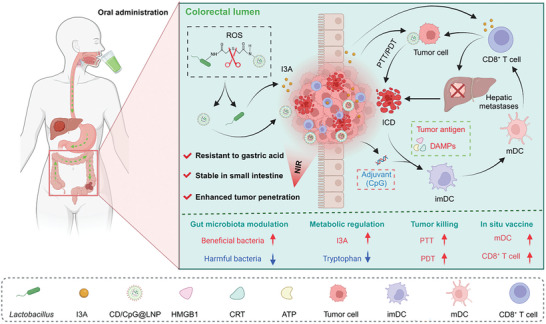
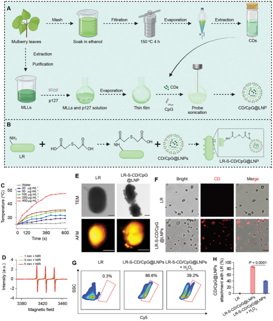
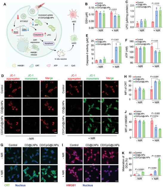
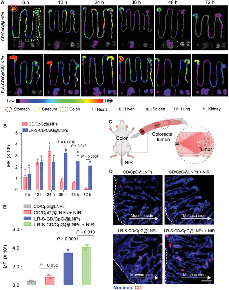
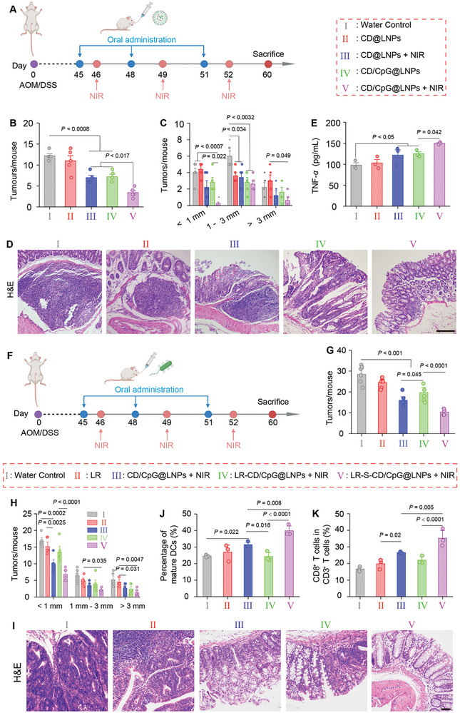
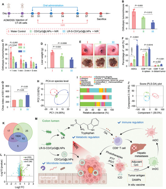
Despite being a groundbreaking approach to treating colorectal cancer (CRC), the efficacy of immunotherapy is significantly compromised by the immunosuppressive tumor microenvironment and dysbiotic intestinal microbiota. Here, leveraging the superior carrying capacity and innate immunity-stimulating property of living bacteria, a nanomedicine-engineered bacterium, LR-S-CD/CpG@LNP, with optical responsiveness, immune-stimulating activity, and the ability to regulate microbiota metabolome is developed. Immunoadjuvant (CpG) and carbon dot (CD) co-loaded plant lipid nanoparticles (CD/CpG@LNPs) are constructed and conjugated to the surface of Limosilactobacillus reuteri (LR) via reactive oxygen species (ROS)-responsive linkers. The inherent photothermal and photodynamic properties of oral CD/CpG@LNPs induce in situ cytotoxic ROS generation and immunogenic cell death of colorectal tumor cells. The generated neoantigens and the released CpG function as a potent in situ vaccine that stimulates the maturation of immature dendritic cells. The mature dendritic cells and metabolites secreted by LR subsequently facilitated the tumor infiltration of cytotoxic T lymphocytes to eradicate colorectal tumors. The further in vivo results demonstrate that the photo-immunotherapy and intestinal microbial metabolite regulation of LR-S-CD/CpG@LNPs collectively suppressed the growth of orthotopic colorectal tumors and their liver metastases, presenting a promising avenue for synergistic treatment of CRC via the oral route.

Keywords: colorectal cancer; engineered bacteria; immunotherapy; oral administration; phototherapy.


Conflict of interest statement

The authors declare no conflict of interest.

Figures

Figure 1
Figure 1
Schematic illustration of oral LR‐S‐CD/CpG@LNPs to achieve tumor accumulation, in situ vaccination, and activation of systemic antitumor immune responses against CRC. After oral administration, nanotherapeutics effectively pass through the upper GIT and accumulate in the deep orthotopic tumor tissues under NIR irradiation. Subsequently, CD/CpG@LNPs induce ICD of tumor cells via PTT/PDT and release abundant autoneoantigens. Neoantigens and CpG are employed in concert to facilitate DC maturation and promote the infiltration of CTLs into the colorectal tumor tissues. Moreover, Lactobacillus enhances the antitumor immune responses by upregulation the level of I3A through the tryptophan metabolic pathway. The synergistic treatment modality of PTT/PDT, in situ vaccination, and I3A not only suppresses orthotopic tumors, but also activates systematic antitumor immunity against distant tumors, increases the abundance of gut beneficial bacteria, and decreases the abundance of harmful bacteria.
Figure 2
Figure 2
Preparation and physicochemical characterization of LR‐S‐CD/CpG@LNPs. Preparation diagram of A) CDs and CD/CpG@LNPs and B) LR‐S‐CD/CpG@LNPs. Insets (A,B) were created with BioRender.com. C) Temperature variations of water and CD suspensions with different CD concentrations as a function of irradiation time with a 660 nm laser (1.0 W cm−2). D) ESR spectra of 1O2 generation of CD/CpG@LNPs upon NIR irradiation with a 660 nm laser (1.0 W cm−2) for different time intervals. E) TEM and AFM images of LR and LR‐S‐CD/CpG@LNPs. Scale bar: 500 nm. F) CLSM images of LR and LR‐S‐CD/CpG@LNPs. The red channel shows CDs. G) FCM analysis of LR‐S‐CD/CpG@LNPs in the absence and presence of H2O2 treatment and H) the corresponding quantitative results (n = 3 independent experiments; *p < 0.05, **p < 0.01, ***p < 0.001, and ****p < 0.0001 by one‐way ANOVA with Tukey's multiple comparison test).
Figure 3
Figure 3
In vitro pro‐apoptosis, ICD, and DC pro‐maturation effects of CD/CpG@LNPs. A) Schematic illustration of the therapeutic mechanism of CD/CpG@LNPs based on phototherapy‐induced apoptosis and ICD, as well as CpG‐regulated DC maturation. Inset (A) was created with BioRender.com. B) The contents of GSH in CT‐26 cells incubated with CD@LNPs and CD/CpG@LNPs at a CD concentration of 2 µg mL−1 with or without NIR irradiation (660 nm, 0.5 W cm−2) for 3 min. C) Quantifying LPO in CT‐26 cells incubated with CD@LNPs and CD/CpG@LNPs at a CD concentration of 2 µg mL−1 with or without NIR irradiation. D) CLSM images of mitochondrial membrane potential changes in CT‐26 cells after different treatments (scare bar: 20 µm). E) Relative enzymatic activities of caspase‐3 in CT‐26 cells incubated with CD@LNPs and CD/CpG@LNPs at a CD concentration of 2 µg mL−1 with or without NIR irradiation. F) Intracellular ATP levels of CT‐26 cells after different treatments. G) CLSM images showing the CRT profiles of CT‐26 cells and H) the corresponding mean fluorescence intensities (MFIs). I) CLSM images showing the HMGB1 profiles of CT‐26 cells and J) the corresponding MFI (scare bar: 20 µm). K) FCM quantification of DC maturation after different treatments (n = 3 independent experiments; *p < 0.05, **p < 0.01, ***p < 0.001, and ****p < 0.0001 by one‐way ANOVA with Tukey's multiple comparison test).
Figure 4
Figure 4
Tumor accumulation and in vivo distribution of CD/CpG@LNPs and LR‐S‐CD/CpG@LNPs with or without NIR irradiation after oral administration. A) Ex vivo fluorescence imaging of the GITs from mice after oral administration of CD/CpG@LNPs and LR‐S‐ CD/CpG@LNPs at 6, 12, 24, 48, and 72 h. B) Following oral administration of CD/CpG@LNPs or LR‐S‐CD/CpG@LNPs at 6, 12, 24, 48, and 72 hours, the corresponding MFIs of the colon were measured. C) Function diagram of the NIR optical fiber for CRC treatment. D) Frozen section imaging of colons from CRC mice receiving oral administration of CD/CpG@LNPs and LR‐S‐CD/CpG@LNPs for 36 h with or without NIR (scare bar: 100 µm). E) MFI of CDs in the colon tissues from various groups (n = 3 independent experiments; *p < 0.05, **p < 0.01, ***p < 0.001, and ****p < 0.0001 by one‐way ANOVA with Tukey's multiple comparison test).
Figure 5
Figure 5
In vivo therapeutic outcomes of various modalities against orthotopic CRC. A) Treatment protocol of different modalities against orthotopic colorectal tumors. B) Total tumor numbers and C) numbers of different‐size tumors per mouse (n = 5 biologically independent experiments; Data were analyzed by one‐way ANOVA with Tukey's multiple comparison test). D) H&E staining of colorectal tumor sections from various mouse groups (scale bar = 100 µm). E) Levels of serum TNF‐α from various mouse groups (n = 3 biologically independent experiments; *p < 0.05, **p < 0.01, ***p < 0.001, and ****p < 0.0001 by one‐way ANOVA with Tukey's multiple comparison test). F) Treatment protocol of LR‐S‐CD/CpG@LNPs (+ NIR) against orthotopic CRC. G) Total tumor numbers and H) numbers of different‐size tumors per mouse (n = 6 biologically independent experiments; *p < 0.05, **p < 0.01, ***p < 0.001, and ****p < 0.0001 by one‐way ANOVA with Tukey's multiple comparison test). I) H&E staining of colorectal tumor sections from various mouse groups (scale bar = 100 µm). J) Percentages of mDCs in the tumor‐draining mesenteric lymph nodes from various mouse groups. K) Percentages of CD8+ T cells in the spleens from various mouse groups (n = 3 biologically independent experiments; *p < 0.05, **p < 0.01, ***p < 0.001, and ****p < 0.0001 by one‐way ANOVA with Tukey's multiple comparison test).
Figure 6
Figure 6
In vivo therapeutic outcomes, intestinal microbiota, and metabolic regulation of various treatment modalities against orthotopic CRC and hepatic metastases. A) Schematic illustration of tumor model establishment and treatment procedures against orthotopic colorectal tumors and hepatic metastases. B) Orthotopic tumor numbers and C) tumor size distributions at the end of treatments. D) Liver weights of all mouse groups at the end of treatments. E) Endoscopic images of colorectal tumors and liver tumor images from various treatment groups. F) Percentages of DCs and CD8+ T cells in the tumor‐draining lymph nodes, spleens, and liver tumors. G) Chao indices of microbes at the ASV levels. H) Principle component analysis (PCA) plots of various mouse groups. I) The relative abundance of the fecal bacterial genus. J) PLS‐DA analysis of the water control, CD/CpG@LNP, and LR‐S‐CD/CpG@LNP groups. K) Venn diagram displaying (comparatively) the differentially expressed metabolites. L) Volcanic maps of intestinal metabolites between the water control and LR‐S‐CD/CpG@LNP groups. M) The therapeutic mechanism of LR‐S‐CD/CpG@LNPs against orthotopic colorectal tumors and hepatic metastases while boosting anti‐tumor immunity. Inset (M) was created with BioRender.com. (n = 4 biologically independent experiments; *p < 0.05, **p < 0.01, ***p < 0.001, and ****p < 0.0001 by one‐way ANOVA with Tukey's multiple comparison test).

Similar articles

Cited by

References

    1. Huang X., Pan J., Xu F., Shao B., Wang Y., Guo X., Zhou S., Adv. Sci. 2021, 8, 2003572. - PMC - PubMed
    1. Guo Y., Chen Y., Liu X., Min J. J., Tan W., Zheng J. H., Cancer Lett. 2020, 469, 102. - PubMed
    1. Chen W., Wang Y., Qin M., Zhang X., Zhang Z., Sun X., Gu Z., ACS Nano 2018, 12, 5995. - PubMed
    1. Gurbatri C. R., Lia I., Vincent R., Coker C., Castro S., Treuting P. M., Hinchliffe T. E., Arpaia N., Danino T., Sci. Transl. Med. 2020, 12, eaax0876. - PMC - PubMed
    1. Chandra D., Jahangir A., Quispe‐Tintaya W., Einstein M. H., Gravekamp C., Br. J. Cancer 2013, 108, 2281. - PMC - PubMed

LinkOut - more resources