. 2024 Dec 31;19(12):e0306197.
doi: 10.1371/journal.pone.0306197. eCollection 2024.

Profiling endogenous airway proteases and antiproteases and modeling proteolytic activation of Influenza HA using in vitro and ex vivo human airway surface liquid samples

Affiliations

Profiling endogenous airway proteases and antiproteases and modeling proteolytic activation of Influenza HA using in vitro and ex vivo human airway surface liquid samples

Stephanie A Brocke et al. PLoS One. .

Abstract

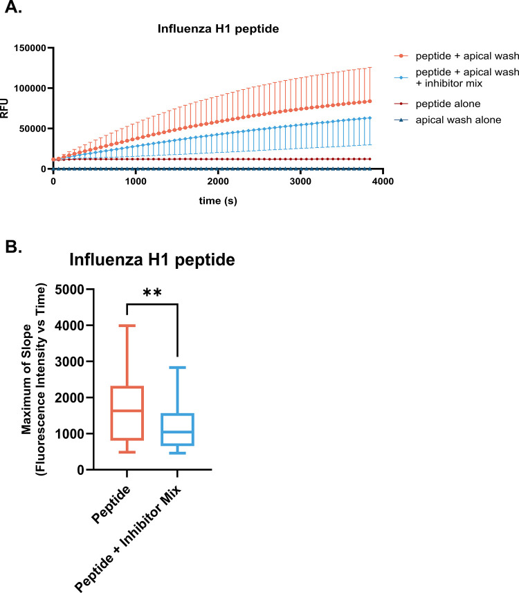
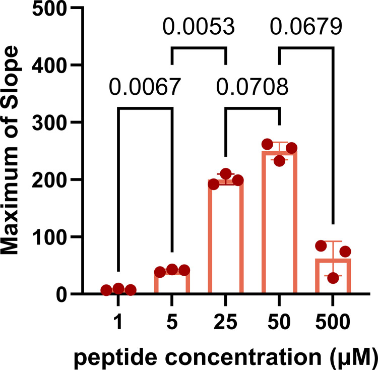
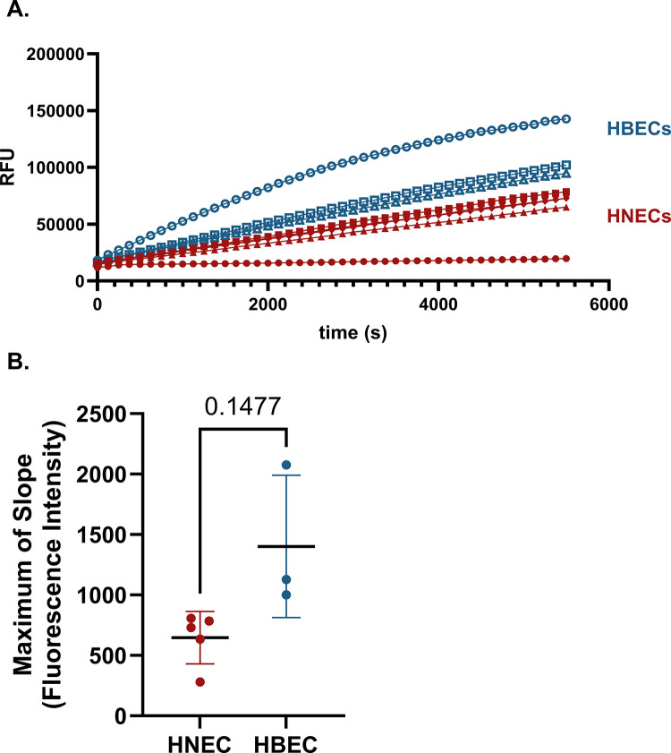
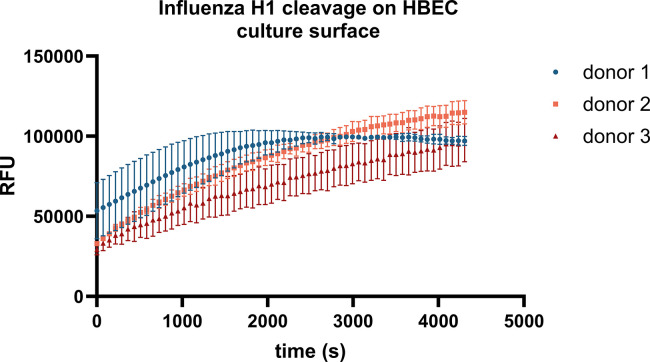
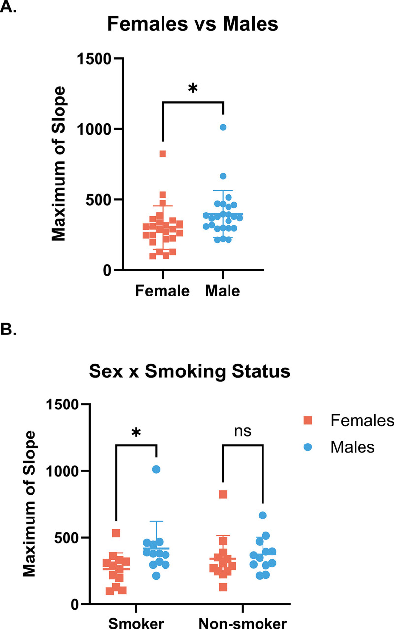
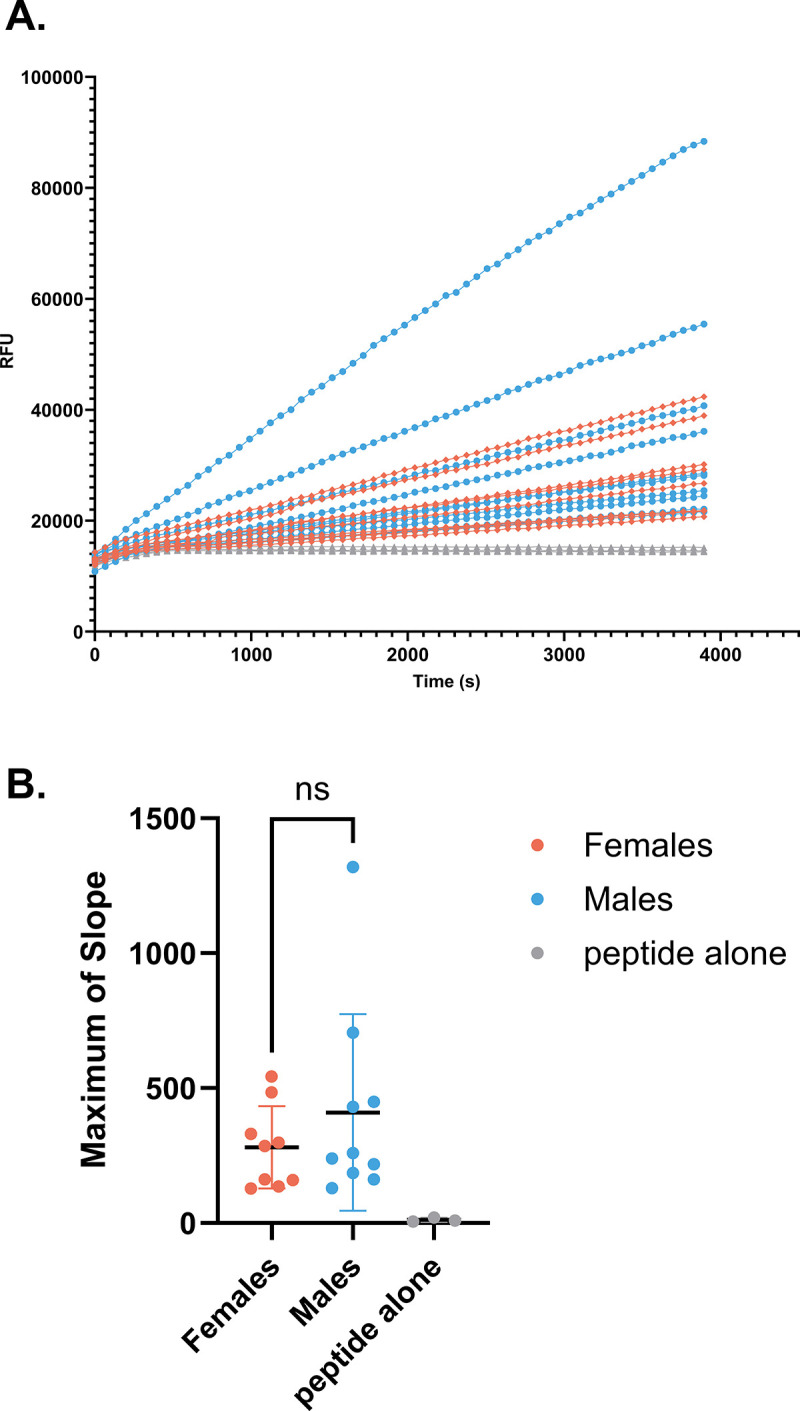
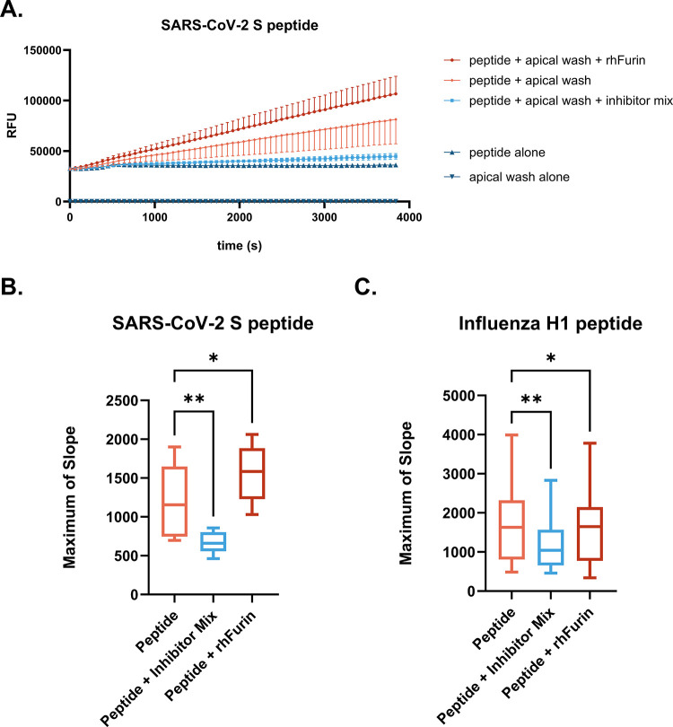
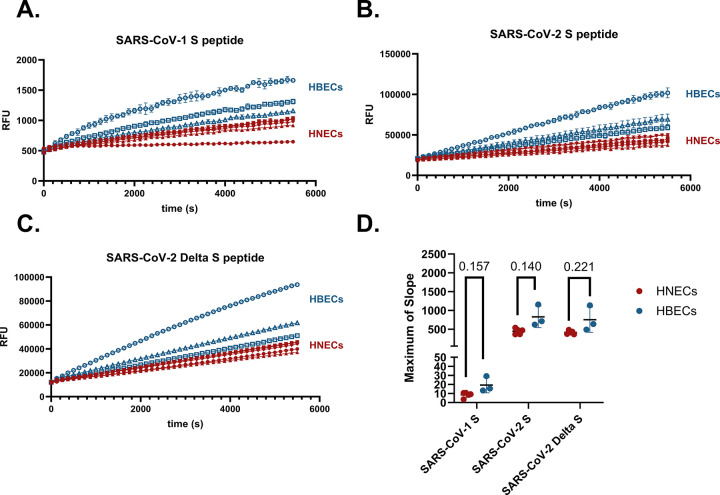
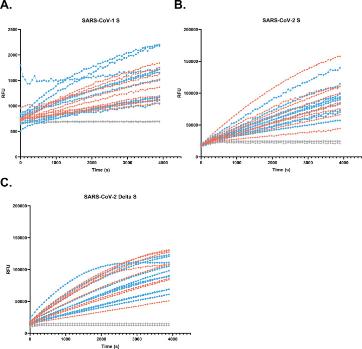
Imbalance of airway proteases and antiproteases has been implicated in diseases such as COPD and environmental exposures including cigarette smoke and ozone. To initiate infection, endogenous proteases are commandeered by respiratory viruses upon encountering the airway epithelium. The airway proteolytic environment likely contains redundant antiproteases and proteases with diverse catalytic mechanisms, however a proteomic profile of these enzymes and inhibitors in airway samples has not been reported. The objective of this study was to first profile extracellular proteases and antiproteases using human airway epithelial cell cultures and ex vivo nasal epithelial lining fluid (NELF) samples. Secondly, we present an optimized method for probing the proteolytic environment of airway surface liquid samples (in vitro and ex vivo) using fluorogenic peptides modeling the cleavage sites of respiratory viruses. We detected 48 proteases in the apical wash of cultured human nasal epithelial cells (HNECs) (n = 6) and 57 in NELF (n = 13) samples from healthy human subjects using mass-spectrometry based proteomics. Additionally, we detected 29 and 48 antiproteases in the HNEC apical washes and NELF, respectively. We observed large interindividual variability in rate of cleavage of an Influenza H1 peptide in the ex vivo clinical samples. Since protease and antiprotease levels have been found to be altered in the airways of smokers, we compared proteolytic cleavage in ex vivo nasal lavage samples from male/female smokers and non-smokers. There was a statistically significant increase in proteolysis of Influenza H1 in NLF from male smokers compared to female smokers. Furthermore, we measured cleavage of the S1/S2 site of SARS-CoV, SARS-CoV-2, and SARS-CoV-2 Delta peptides in various airway samples, suggesting the method could be used for other viruses of public health relevance. This assay presents a direct and efficient method of evaluating the proteolytic environment of human airway samples in assessment of therapeutic treatment, exposure, or underlying disease.


Conflict of interest statement

The authors have declared that no competing interests exist.

Figures

Fig 1
Fig 1. Proteases in HNEC apical wash cleave Influenza H1.
HNECs from n = 9 (5M, 4F) donors were cultured at air liquid interface. Apical washes from differentiated cultures were assayed for proteolytic activity toward the influenza H1 peptide by mixing each sample with 50 μM of the peptide and measuring fluorescence intensity over time. A) The mean change in relative fluorescence units (RFU) versus time (s) for all donors is shown in orange. Each donor was assayed in triplicate. Addition of a protease inhibitor mix on rate of cleavage is indicated by the light blue line. Controls include the peptide at 50 μM without addition of HNEC apical wash (red) and HNEC apical wash alone, without the peptide (dark blue). Samples were collected 4 d since the prior apical wash. B) Maximum of slope of the cleavage reaction is plotted and a paired t-test was used to evaluate differences between groups; ** indicates p<0.01.
Fig 2
Fig 2. Rate of cleavage increases with substrate concentration up to 50 μM.
Maximum rates of cleavage of Influenza H1 peptide at a range of concentrations from 1–500 μM tested with apical wash samples from n = 3 (2M, 1F) HNEC donors. A repeated measures one-way ANOVA with Bonferroni’s post hoc test was used to determine differences between groups, p≤0.05.
Fig 3
Fig 3. Comparison of Influenza H1 cleavage by proteases in nasal vs bronchial samples.
A) HNEC and HBEC cultures grown at ALI were apically washed. Samples were then mixed with an influenza H1 IQF peptide and fluorescence intensity in each sample over time was measured in a microplate reader. Rates of cleavage of Influenza H1 by proteases in apical wash samples from n = 5 (3M, 2F) HNEC donors and n = 3 (2M, 1F) HBEC donors are shown, each line representing a single donor averaging three technical replicates with standard deviation. HBECs are shown in blue and HNECs are shown in red. All samples were collected 7 d from the prior apical wash. B) Maximum of slope of the cleavage reaction is plotted. A Welch’s unequal variances t-test was used to test for difference between groups.
Fig 4
Fig 4. Proteolytic activity measured directly on the apical surface of ALI cultures.
Proteolytic activity was measured on HBEC cultures at ALI. A 50 μM solution of Influenza H1 peptide in 100 μl of total volume was added to the apical surface of duplicate cultures from n = 3 (2M, 1F) donors. The prior apical wash of the cultures occurred 72h prior. Change in fluorescence intensity over time (s) on the surface of the cultures was then measured on a plate reader.
Fig 5
Fig 5. Differences in cleavage of Influenza H1 by proteases in NLF from male and female smokers.
NLF samples were collected from (n = 48) smoking and non-smoking adults. The Influenza H1 peptide solution (50 μM) was then mixed with the samples and fluorescence intensity was measured on a plate reader. No outliers were identified by the ROUT (Q = 1%) method. A) Sex difference between n = 24 Male and n = 24 Female donors when the data are aggregated by smoking status (unpaired t-test, p≤0.05). B) Separating by smoking status demonstrates greater cleavage of the Influenza H1 peptide in samples from male smokers compared to female smokers, with no statistically significant difference between non-smokers (2-way ANOVA with Bonferroni‘s post hoc test, p≤0.05). In both plots, mean with standard deviation is shown.
Fig 6
Fig 6. Proteases in NELF cleave Influenza H1.
NELF samples were collected and mixed with the IQF Influenza peptide solution. Change in fluorescence intensity over time was then measured with a plate reader. A) Cleavage of the Influenza H1 peptide by proteases in NELF samples from n = 19 donors, males in blue (n = 10), and females in orange (n = 9). Triplicates of a peptide-only control (i.e. no NELF) are in light gray. B) Maximum rate of cleavage for all samples. Maximums of slope were calculated for data in the range of time = 480-3894s. Welch’s unequal variance t-test was used to evaluate difference in maximum slope between sexes.
Fig 7
Fig 7. Cleavage of SARS-CoV-2 S IQF peptide by apical wash samples from HNECs.
Cultures from n = 9 (5M, 4F) HNEC donors were apically washed. At the time of assay, samples were then mixed with the 50μM SARS-CoV-2 IQF peptide solution and the cleavage reaction was observed in a plate reader. A) Change in RFU vs time is shown in orange. The addition of a mixture of protease inhibitors (light blue) or rhFurin (red) on rate of cleavage is also shown. Controls (peptide with no sample or HNEC samples with no peptide) are in dark blue. Samples were collected 4 d since the prior apical wash. B) Maximum rates of reaction for SARS-CoV-2 S and C) for Influenza H1. Differences between groups were detected by repeated measures one-way ANOVA with Dunnett‘s post hoc test; * p≤0.05, ** p≤0.01.
Fig 8
Fig 8. Proteases in HNEC and HBEC apical washes cleave SARS coronavirus peptides.
Rates of cleavage of SARS S1/S2 IQF peptides by apical wash samples from n = 5 (3M, 2F) HNEC donors (in red) and n = 3 (2M, 1F) HBEC donors (in blue). Cleavage of A) SARS-CoV-1 B) SARS-CoV-2, and C) SARS-CoV-2 Delta. All samples were collected 7 d from the prior apical wash. Each line represents the mean of three technical replicates with standard deviation. D) Differences in maximum of slope between HBECs and HNECs for each peptide were tested with individual Welch’s unequal variance t-tests.
Fig 9
Fig 9. Proteases in NELF cleave SARS coronavirus peptides.
Rate of cleavage of the SARS coronavirus peptides (50 μM) by proteases in NELF samples from n = 19 donors, males in blue (n = 10), and females in orange (n = 9). Peptide-only controls are shown in light gray. Cleavage of SARS-CoV-1 is shown in (A), SARS-CoV-2 in (B) and SARS-CoV-2 Delta in (C).

Similar articles

  • The effect of sample site and collection procedure on identification of SARS-CoV-2 infection.
    Davenport C, Arevalo-Rodriguez I, Mateos-Haro M, Berhane S, Dinnes J, Spijker R, Buitrago-Garcia D, Ciapponi A, Takwoingi Y, Deeks JJ, Emperador D, Leeflang MMG, Van den Bruel A; Cochrane COVID-19 Diagnostic Test Accuracy Group. Davenport C, et al. Cochrane Database Syst Rev. 2024 Dec 16;12(12):CD014780. doi: 10.1002/14651858.CD014780. Cochrane Database Syst Rev. 2024. PMID: 39679851 Free PMC article.
  • Physical interventions to interrupt or reduce the spread of respiratory viruses.
    Jefferson T, Dooley L, Ferroni E, Al-Ansary LA, van Driel ML, Bawazeer GA, Jones MA, Hoffmann TC, Clark J, Beller EM, Glasziou PP, Conly JM. Jefferson T, et al. Cochrane Database Syst Rev. 2023 Jan 30;1(1):CD006207. doi: 10.1002/14651858.CD006207.pub6. Cochrane Database Syst Rev. 2023. PMID: 36715243 Free PMC article.
  • Rapid, point-of-care antigen tests for diagnosis of SARS-CoV-2 infection.
    Dinnes J, Sharma P, Berhane S, van Wyk SS, Nyaaba N, Domen J, Taylor M, Cunningham J, Davenport C, Dittrich S, Emperador D, Hooft L, Leeflang MM, McInnes MD, Spijker R, Verbakel JY, Takwoingi Y, Taylor-Phillips S, Van den Bruel A, Deeks JJ; Cochrane COVID-19 Diagnostic Test Accuracy Group. Dinnes J, et al. Cochrane Database Syst Rev. 2022 Jul 22;7(7):CD013705. doi: 10.1002/14651858.CD013705.pub3. Cochrane Database Syst Rev. 2022. PMID: 35866452 Free PMC article.
  • Type I and Type III Interferons Restrict SARS-CoV-2 Infection of Human Airway Epithelial Cultures.
    Vanderheiden A, Ralfs P, Chirkova T, Upadhyay AA, Zimmerman MG, Bedoya S, Aoued H, Tharp GM, Pellegrini KL, Manfredi C, Sorscher E, Mainou B, Lobby JL, Kohlmeier JE, Lowen AC, Shi PY, Menachery VD, Anderson LJ, Grakoui A, Bosinger SE, Suthar MS. Vanderheiden A, et al. J Virol. 2020 Sep 15;94(19):e00985-20. doi: 10.1128/JVI.00985-20. Print 2020 Sep 15. J Virol. 2020. PMID: 32699094 Free PMC article.
  • Antibody tests for identification of current and past infection with SARS-CoV-2.
    Fox T, Geppert J, Dinnes J, Scandrett K, Bigio J, Sulis G, Hettiarachchi D, Mathangasinghe Y, Weeratunga P, Wickramasinghe D, Bergman H, Buckley BS, Probyn K, Sguassero Y, Davenport C, Cunningham J, Dittrich S, Emperador D, Hooft L, Leeflang MM, McInnes MD, Spijker R, Struyf T, Van den Bruel A, Verbakel JY, Takwoingi Y, Taylor-Phillips S, Deeks JJ; Cochrane COVID-19 Diagnostic Test Accuracy Group. Fox T, et al. Cochrane Database Syst Rev. 2022 Nov 17;11(11):CD013652. doi: 10.1002/14651858.CD013652.pub2. Cochrane Database Syst Rev. 2022. PMID: 36394900 Free PMC article.

References

    1. Rawlings ND, Barrett AJ, Thomas PD, Huang X, Bateman A, Finn RD. The MEROPS database of proteolytic enzymes, their substrates and inhibitors in 2017 and a comparison with peptidases in the PANTHER database. Nucleic Acids Research. 2017;46(D1):D624–D32. - PMC - PubMed
    1. Fugère M, Day R. Cutting back on pro-protein convertases: the latest approaches to pharmacological inhibition. Trends Pharmacol Sci. 2005;26(6):294–301. doi: 10.1016/j.tips.2005.04.006 - DOI - PMC - PubMed
    1. Antalis T.M., Buzza M.S. Extracellular: Plasma Membrane Proteases—Serine Proteases. Encyclopedia of Cell Biology. 2016:650–60.
    1. Sepper R, Konttinen YT, Ingman T, Sorsa T. Presence, activities, and molecular forms of cathepsin G, elastase,α1-antitrypsin, andα1-antichymotrypsin in bronchiectasis. Journal of Clinical Immunology. 1995;15(1):27–34. - PubMed
    1. S Yasuoka TO, Kawano S, Tsuchihashi S, Ogawara M, Masuda K, Yamaoka K, et al.. Purification, characterization, and localization of a novel trypsin-like protease found in the human airway. American Journal of Respiratory Cell and Molecular Biology. 1997;16(3):300–8. doi: 10.1165/ajrcmb.16.3.9070615 - DOI - PubMed

Substances

LinkOut - more resources