. 2024 Dec 31;19(12):e0308428.
doi: 10.1371/journal.pone.0308428. eCollection 2024.

Deciphering the interactome of Ataxin-2 and TDP-43 in iPSC-derived neurons for potential ALS targets

Affiliations

Deciphering the interactome of Ataxin-2 and TDP-43 in iPSC-derived neurons for potential ALS targets

Yuan Tian et al. PLoS One. .

Abstract

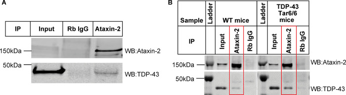
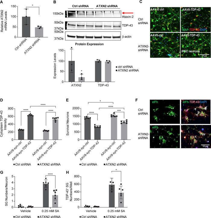
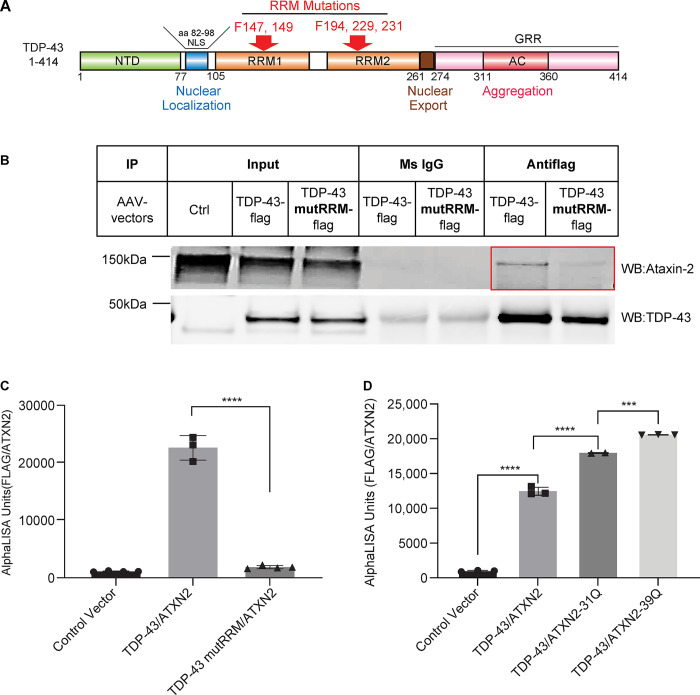
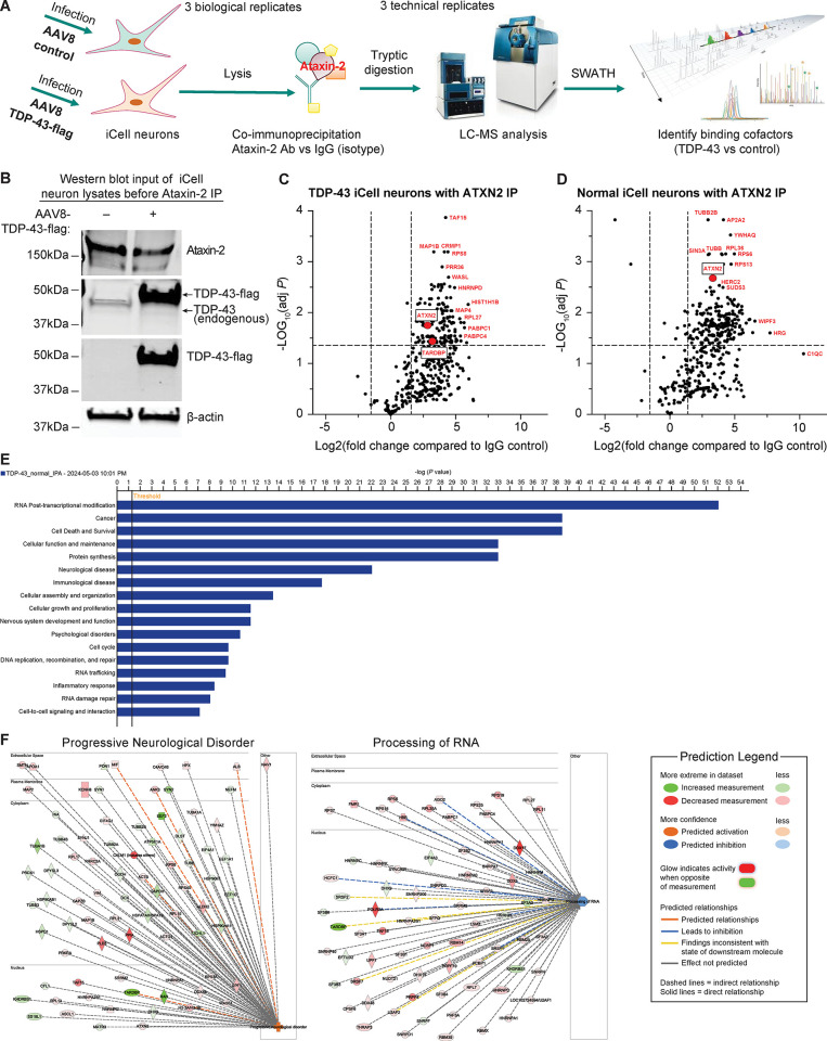
Ataxin-2 is a protein containing a polyQ extension and intermediate length of polyQ extensions increases the risk of Amyotrophic Lateral Sclerosis (ALS). Down-regulation of Ataxin-2 has been shown to mitigate TDP-43 proteinopathy in ALS models. To identify alternative therapeutic targets that can mitigate TDP-43 toxicity, we examined the interaction between Ataxin-2 and TDP-43. Co-immunoprecipitation demonstrated that Ataxin-2 and TDP-43 interact, that their interaction is mediated through the RNA recognition motif (RRM) of TDP-43, and knocking down Ataxin-2 or mutating the RRM domains rescued TDP-43 toxicity in an iPSC-derived neuronal model with TDP-43 overexpression. To decipher the Ataxin-2 and TDP-43 interactome, we used co-immunoprecipitation followed by mass spectrometry to identify proteins that interacted with Ataxin-2 and TDP-43 under conditions of endogenous or overexpressed TDP-43 in iPSC-derived neurons. Multiple interactome proteins were differentially regulated by TDP-43 overexpression and toxicity, including those involved in RNA regulation, cell survival, cytoskeleton reorganization, protein modification, and diseases. Interestingly, the RNA-binding protein (RBP), TAF15 which has been implicated in ALS was identified as a strong binder of Ataxin-2 in the condition of TDP-43 overexpression. Together, this study provides a comprehensive annotation of the Ataxin-2 and TDP-43 interactome and identifies potential therapeutic pathways and targets that could be modulated to alleviate Ataxin-2 and TDP-43 interaction-induced toxicity in ALS.


Conflict of interest statement

All authors were employees of Merck & Co., Inc., Rahway, NJ, USA at the time of this study. Employment does not alter authors’ adherence to the journal’s policies on conflicts of interest or sharing data and materials.

Figures

Fig 1
Fig 1. Ataxin-2 co-immunoprecipitated with TDP-43 in iPSC-derived neurons and mouse brain tissue.
Ataxin-2 co-immunoprecipitated with TDP-43 in human iCell neurons (A) and mouse brain tissue (B). The iCell neurons were infected with AAV8-syn-TDP-43, and the lysate was immunoprecipitated with either IgG or Ataxin-2 antibody. The elutes were followed by Western blot with Ataxin-2 and TDP-43 antibodies. WT and Tar6/Tar6 mouse brain lysates were immunoprecipitated with IgG or Ataxin-2 antibody, respectively. The elutes were followed by Western blot with Ataxin-2 and TDP-43 antibodies.
Fig 2
Fig 2. ATXN2 knock-down alleviated TDP-43 overexpression-induced neuronal loss and stress granule formation in iPSC-derived GABA neurons.
Human iPSC-derived GABAergic neurons that were infected with LV-shRNA ctrl or LV-shRNA ATXN2 were then transduced with AAV-control or AAV-TDP-43. (A) Cell lysates were analyzed by qPCR to confirm the ATXN2 mRNA knockdown (n = 3, mean ± SD, * p<0.05, compared to the control group, two-sample t-test). (B) Western blot was performed with anti-Ataxin-2 and TDP-43 antibodies (upper panel) for protein expression levels. The images were quantified and normalized to LV-shRNA-ctrl treated cells as 100% (lower panel) (n = 3, mean ± SD, * p<0.05, compared to the control group, two-sample t-test). (C) ICC was performed with anti-TDP-43 antibody (red) and anti-Map-2 antibody (green) to visualize neuronal bodies and neurites. Nuclei were stained by Hoechst (blue). (D) High-content imaging quantification of cytoplasm TDP-43 (RU per cell). (E) Survival neurons by MAP-2 staining (per well) (n = 3, mean ± SD * p<0.05, ** p<0.01, *** p<0.001, **** p<0.0001 two-way ANOVA followed by Šidák’s multiple comparisons test). (F) High-content imaging of ICC with anti-TDP-43 antibody (red) and anti-eIFh antibody (green) to visualize stress granules. Nuclei were stained by Hoechst (blue). The iPSC-derived GABA neurons that were infected with LV-shRNA-ctrl and LV-shRNA-ATXN2 then infected with AAV-TDP-43 before treated with Sodium Arsenite (SA) for 2hrs to induce stress granule formation. Arrows indicate stress granules containing TDP43. (G) High-content imaging quantification of eIFh+ stress granule numbers per neuron (n = 9/group, 2 independent experiments, mean ± SD, ***p<0.0001, two-way ANOVA followed by Šidák’s multiple comparisons test). (H) High-content imaging quantification of TDP-43 positive stress granule numbers per well, which is defined as stress granules that have a well-defined TDP-43 staining overlap (n = 9/group, 2 independent experiments, mean ± SD, ****p<0.0001, two-way ANOVA followed by Šidák’s multiple comparisons test).
Fig 3
Fig 3. Ataxin-2 interacted with TDP-43 via the RRM domains and polyQ extension enhanced the interaction.
(A) Schematic representation of TDP-43 protein and the RRM mutations that abolish its interaction with RNA binding proteins. (B) Co-immunoprecipitation and western blot of TDP-43 and TDP-43 RRM mutant with Ataxin-2. Human iPSC-derived GABA neurons were infected with scAAV8-Syn-TDP-43-flag or scAAV8-Syn-TDP-43mutRRM-flag, and the lysates were immunoprecipitated with Flag antibody. Western blot was performed with Ataxin-2 and TDP-43 antibodies. (C) Proximity AlphaLISA for the interaction between TDP-43 and TDP-43mutRRM with Ataxin-2. HEK293 cells were transfected with either empty vector, or vectors co-overexpressing ATXN2 and TDP-43-flag or ATXN2 and TDP-43mutRRM-flag, respectively. Cell lysates were analyzed by proximity AlphaLISA (n = 3, mean ± SD * p<0.05, ** p<0.01, *** p<0.001, **** p<0.0001 one-way ANOVA followed by Tukey’s multiple comparison test). (D) Proximity AlphaLISA for the interaction between either Ataxin-2, or Ataxin-2-31Q, or Ataxin-2-39Q and TDP-43. HEK293 cells were transfected with either empty vector, or vectors overexpressing TDP-43-flag and ATXN2, TDP-43-flag and ATXN2-31Q or TDP-43-flag and ATXN2-39Q, respectively. Cell lysates were analyzed by proximity AlphaLISA (n = 3, mean ± SD * p<0.05, ** p<0.01, *** p<0.001, **** p<0.0001 one-way ANOVA followed by Tukey’s multiple comparison test).
Fig 4
Fig 4. TDP-43 RRM mutant abolished TDP-43-induced toxicity in iPSC-derived neurons.
(A) ICC of human iPSC-derived GABA neurons that were infected with AAV-control (upper panel), AAV-TDP-43 (middle panel) or AAV-TDP-43mutRRM (lower panel). Immunostaining was performed with anti-TDP-43 antibody (red) and anti-Map-2 antibody (green) to visualize neuronal bodies and neurites. Nuclei were stained by Hoechst (blue). (B) High-content imaging quantification of MAP-2 positive cells per well (n = 3, mean ± SD * p<0.05, ** p<0.01, *** p<0.001, **** p<0.0001 one-way ANOVA followed by Tukey’s multiple comparison test). (C) High-content imaging quantification Caspase 3 positive cells as percentage to total cells per well (n = 3, mean ± SD * p<0.05, ** p<0.01, *** p<0.001, **** p<0.0001 one-way ANOVA followed by Tukey’s multiple comparison test). (D) High-content imaging quantification of cytoplasm TDP-43 intensity per well of RU (n = 3, mean ± SD * p<0.05, ** p<0.01, *** p<0.001, **** p<0.0001 one-way ANOVA followed by Tukey’s multiple comparison test).Co-Immunoprecipitation followed by Mass Spectrometry (IP-MS) identified Ataxin-2 interactome in iPSC derived neurons.
Fig 5
Fig 5. IP-MS identified ATXN2 interactome in the conditions of endogenous (Normal) or TDP-43 overexpression (TDP-43) in iPSC-derived GABA neurons.
(A) Schematic representation of the IP-MS procedure. Human iPSC-derived GABA neurons were infected with either AAV-ctrl or AAV-TDP-43, and the lysate was immunoprecipitated with either isotype IgG control or Ataxin-2 antibody with 3 biological replicates. The elutes were followed by tryptic digestion and LC-SWATH MS analysis with 3 technical replicates to identify binding cofactors of TDP-43 overexpression vs control (endogenous TDP-43). (B) Western blot of the input cell lysates before Ataxin-2 IP. Aliquots of the cell lysates of either AAV-ctrl or AAV-TDP-43 infected neurons were analyzed by Western blot with Ataxin-2 and TDP-43 antibodies. (C, D) Volcano plot of all interacting proteins identified from TDP-43 overexpression (TDP-43) vs endogenous expression (Normal). They were shown with Fold Change = Mean Ave of Ataxin-2 (3 bioreplicates) over Mean Ave IgG (3 bioreplicates). Horizontal dotted line for Adj. p-value = 0.05, Welch T-Test (assume unequal variances), and vertical dotted lines for Fold Change = 3 or -3. (E) IPA analysis of the canonical pathways, biological functions, and disease pathways that the hits involved. The line bar represents the threshold of significance (p = 0.05). (F) The interaction networks of the hits functionally related to progressive neurological disorders and progressing of RNA.
Fig 6
Fig 6. Interactome hits of TDP-43 overexpression vs normal.
(A) Volcano plot of hit overlay of fold changes for TDP-43 overexpression over Normal (endogenous). Proteins highlighted in red have a fold change>2 compared to normal; Welch T-Test (assume unequal variances), Adj. p-value<0.05. Horizontal dotted line for Adj. p-value = 0.05, Welch T-Test (assume unequal variances), and vertical dotted lines for Fold Change = 2 or -2. (B) Venn plot summary of hits distinct to TDP-43 (overexpression) vs Normal (endogenous).

Similar articles

References

    1. Ou SH, Wu F, Harrich D, García-Martínez LF, Gaynor RB. Cloning and characterization of a novel cellular protein, TDP-43, that binds to human immunodeficiency virus type 1 TAR DNA sequence motifs. J Virol. 1995;69(6):3584–96. doi: 10.1128/JVI.69.6.3584-3596.1995 - DOI - PMC - PubMed
    1. Neumann M, Sampathu DM, Kwong LK, Truax AC, Micsenyi MC, Chou TT, et al.. Ubiquitinated TDP-43 in frontotemporal lobar degeneration and amyotrophic lateral sclerosis. Science. 2006;314(5796):130–3. doi: 10.1126/science.1134108 - DOI - PubMed
    1. Imbert G, Saudou F, Yvert G, Devys D, Trottier Y, Garnier JM, et al.. Cloning of the gene for spinocerebellar ataxia 2 reveals a locus with high sensitivity to expanded CAG/glutamine repeats. Nat Genet. 1996;14(3):285–91. doi: 10.1038/ng1196-285 - DOI - PubMed
    1. Pulst SM, Nechiporuk A, Nechiporuk T, Gispert S, Chen XN, Lopes-Cendes I, et al.. Moderate expansion of a normally biallelic trinucleotide repeat in spinocerebellar ataxia type 2. Nat Genet. 1996;14(3):269–76. doi: 10.1038/ng1196-269 - DOI - PubMed
    1. Sanpei K, Takano H, Igarashi S, Sato T, Oyake M, Sasaki H, et al.. Identification of the spinocerebellar ataxia type 2 gene using a direct identification of repeat expansion and cloning technique, DIRECT. Nat Genet. 1996;14(3):277–84. doi: 10.1038/ng1196-277 - DOI - PubMed

MeSH terms