. 2024 Dec 31;19(12):e0315510.
doi: 10.1371/journal.pone.0315510. eCollection 2024.

Mechanism of connexin channel inhibition by mefloquine and 2-aminoethoxydiphenyl borate

Affiliations

Mechanism of connexin channel inhibition by mefloquine and 2-aminoethoxydiphenyl borate

Pia Lavriha et al. PLoS One. .

Abstract

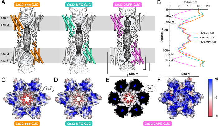
Gap junction intercellular communication (GJIC) between two adjacent cells involves direct exchange of cytosolic ions and small molecules via connexin gap junction channels (GJCs). Connexin GJCs have emerged as drug targets, with small molecule connexin inhibitors considered a viable therapeutic strategy in several diseases. The molecular mechanisms of GJC inhibition by known small molecule connexin inhibitors remain unknown, preventing the development of more potent and connexin-specific therapeutics. Here we show that two GJC inhibitors, mefloquine (MFQ) and 2-aminoethoxydiphenyl borate (2APB) bind to Cx32 and block dye permeation across Cx32 hemichannels (HCs) and GJCs. Cryo-EM analysis shows that 2APB binds to "site A", close to the N-terminal gating helix of Cx32 GJC, restricting the entrance to the channel pore. In contrast, MFQ binds to a distinct "site M", deeply buried within the pore. MFQ binding to this site modifies the electrostatic properties of Cx32 pore. Mutagenesis of V37, a key residue located in the site M, renders Cx32 HCs and GJCs insensitive to MFQ-mediated inhibition. Moreover, our cryo-EM analysis, mutagenesis and activity assays show that MFQ targets the M site in Cx43 GJC similarly to Cx32. Taken together, our results point to a conserved inhibitor binding site in connexin channels, opening a new route for development of specific drugs targeting connexins.


Conflict of interest statement

The authors have declared that no competing interests exist.

Figures

Fig 1
Fig 1. Analysis of Cx32 channel inhibition by MFQ and 2APB.
(A) Schematic representation of connexin inhibition by MFQ and 2APB. (B) Dye uptake of Cx32 HC and mock-transfected control cells upon treatment with MFQ or DMSO (drug solvent) (Cx32: MFQ, n = 1183; DMSO, n = 1109; Control: MFQ, n = 1047; DMSO, n = 1016). The HC dye-uptake values were compared using Games-Howell’s multiple comparisons test: ****—P < 0.0001, ***—P < 0.001, **—P < 0.01, *—P < 0.05, ns–P > 0.05. n–number of measured cells; all experiments were performed in experimental triplicates. (C) GJC permeability in Cx32 and mock-transfected control cells upon MFQ, or DMSO treatment (Cx32: DMSO, n = 12; MFQ, n = 16; Control: DMSO, n = 7; MFQ, n = 13). (D) Cx32 tryptophan fluorescence quenching upon MFQ treatment (n = 3). (E) Dye uptake of Cx32 HC and mock-transfected control cells upon treatment with 2APB or DMSO (Cx32: 2APB, n = 911; DMSO, n = 1109; Control: 2APB, n = 1047; DMSO, n = 1016). n–number of measured cells; all experiments were performed in experimental triplicates. (F) GJC permeability for Cx32 and mock-transfected control upon 2APB, or DMSO treatment (Cx32: DMSO, n = 12; 2APB, n = 15; Control: DMSO, n = 7; 2APB, n = 12). (G) Spectral shift of tryptophan fluorescence upon 2APB titration (n = 3). The values (%) on the right side in D and G indicate the distribution of the calculated low and high affinity binding sites. All data is represented as mean ± SEM. (H) Side views of cryo-EM map and model of Cx32 GJC in complex with MFQ. (I) Same as H, for Cx32-2APB (level = 4σ). Additional densities in H and I, interpreted to be MFQ and 2APB respectively, are illustrated with colored features. TMD–transmembrane domain; ECD–extracellular domain; In–intracellular side; Out–extracellular side.
Fig 2
Fig 2. Locations of the MFQ and 2APB binding sites in the Cx32 GJC.
(A) Schematic representation of putative MFQ and 2APB binding sites A and M in the Cx32 GJC. (B-C) Views of the Cx32-MFQ (B) and Cx32-2APB maps (C), contoured at 4σ. (D-E) A comparison of the densities at sites A and M in Cx32-apo GJC and Cx32-MFQ GJC, Cx32-2APB GJC (D) and Cx32-apo HC (E) (all maps contoured at 3σ). TM–transmembrane helix, NTH–N-terminal helix.
Fig 3
Fig 3. Molecular details of the inhibitor binding sites in Cx32.
(A) Locations of site A and M are shown with the boxes (right). Left: site A. Residues (side chains shown as sticks) constituting the site A, within 4 Å of the bound 2APB molecule, are shown with white labels. The sterol molecule (green) is located in close vicinity of the site A. (B-C) Site M. Side chains of the residues in close proximity of the MFQ (B) or 2APB (C) are shown as sticks. Purple labels indicate residues linked to CMT1X disease. (D-G) GJC permeability of V37W and V37I Cx32 mutants upon treatment with MFQ (D, F) and 2APB (E, G). The GJC activity values are shown as mean ± SEM; for D, n = 20 (DMSO) and n = 18 (MFQ); for E, n = 20 (DMSO) and n = 16 (2APB); for F, n = 17 (DMSO) and n = 19 (MFQ); for G, n = 17 (DMSO) and n = 20 (2APB).
Fig 4
Fig 4. Effect of drug binding to site A and M on Cx32 GJC pore properties.
(A) Diffusive pathways for the solutes of Cx32 and Cx32 in complex with MFQ or 2APB, calculated using HOLE. (B) Pore radii along the pore, calculated using HOLE (shown in A). (C-D) Electrostatic surface potential representations of Cx32-apo GJC (C), Cx32-MFQ (D). (E) The clipped view of Cx32-2APB revealing the site M. (F) The unclipped view of Cx32-2APB (as in C and D).
Fig 5
Fig 5. Functional and structural analysis of MFQ effect on Cx43 GJC.
(A) A view of the cryo-EM map and model of Cx43 GJC in complex with MFQ. (B) Cryo-EM map of Cx43-MFQ, viewed from the cytosolic side; a clipped view, excluding the NTH density is shown. (C) Comparison of site M density of Cx43-apo GJC and Cx43-MFQ GJC (contoured at 3.3σ). (D) GJC permeability of WT Cx43 (top) and G38W (bottom) with or without MFQ treatment (n = 15). All data in D are represented as mean ± SEM. Inset: A G38W mutation sterically hinders MFQ binding, with the side chain of W38 clashing with the ligand. (E) Comparison of electrostatic surface potential of Cx43-apo (PDB ID: 7Z1T) and Cx43-MFQ GJCs.
Fig 6
Fig 6. Mechanism of connexin channel inhibition via sites A and M.
(A) In the absence of the inhibitors, the connexin GJCs allow free passage of solutes (<1.5 kDa). (B) Binding of MFQ to site M introduces and electrostatic barrier in the pore, reducing GJCs permeability. (C) 2APB binds to site M and to site A, causing NTH rearrangement and constriction of the pore entrance.

Similar articles

References

    1. Dbouk HA, Mroue RM, El-Sabban ME, Talhouk RS. Connexins: a myriad of functions extending beyond assembly of gap junction channels. Cell Commun Signal. 2009;7:4. Epub 20090312. doi: 10.1186/1478-811X-7-4 ; PubMed Central PMCID: PMC2660342. - DOI - PMC - PubMed
    1. Nielsen MS, Axelsen LN, Sorgen PL, Verma V, Delmar M, Holstein-Rathlou NH. Gap junctions. Compr Physiol. 2012;2(3):1981–2035. doi: 10.1002/cphy.c110051 ; PubMed Central PMCID: PMC3821273. - DOI - PMC - PubMed
    1. Totland MZ, Rasmussen NL, Knudsen LM, Leithe E. Regulation of gap junction intercellular communication by connexin ubiquitination: physiological and pathophysiological implications. Cell Mol Life Sci. 2020;77(4):573–91. Epub 20190909. doi: 10.1007/s00018-019-03285-0 ; PubMed Central PMCID: PMC7040059. - DOI - PMC - PubMed
    1. Kurtenbach S, Kurtenbach S, Zoidl G. Gap junction modulation and its implications for heart function. Front Physiol. 2014;5:82. Epub 20140227. doi: 10.3389/fphys.2014.00082 ; PubMed Central PMCID: PMC3936571. - DOI - PMC - PubMed
    1. Bortolozzi M. What’s the Function of Connexin 32 in the Peripheral Nervous System? Front Mol Neurosci. 2018;11:227. Epub 20180710. doi: 10.3389/fnmol.2018.00227 ; PubMed Central PMCID: PMC6048289. - DOI - PMC - PubMed

LinkOut - more resources