. 2025 Jan 7;122(1):e2415250121.
doi: 10.1073/pnas.2415250121. Epub 2024 Dec 31.

Topological confinement by a membrane anchor suppresses phase separation into protein aggregates: Implications for prion diseases

Affiliations

Topological confinement by a membrane anchor suppresses phase separation into protein aggregates: Implications for prion diseases

Kalpshree Gogte et al. Proc Natl Acad Sci U S A. .

Abstract

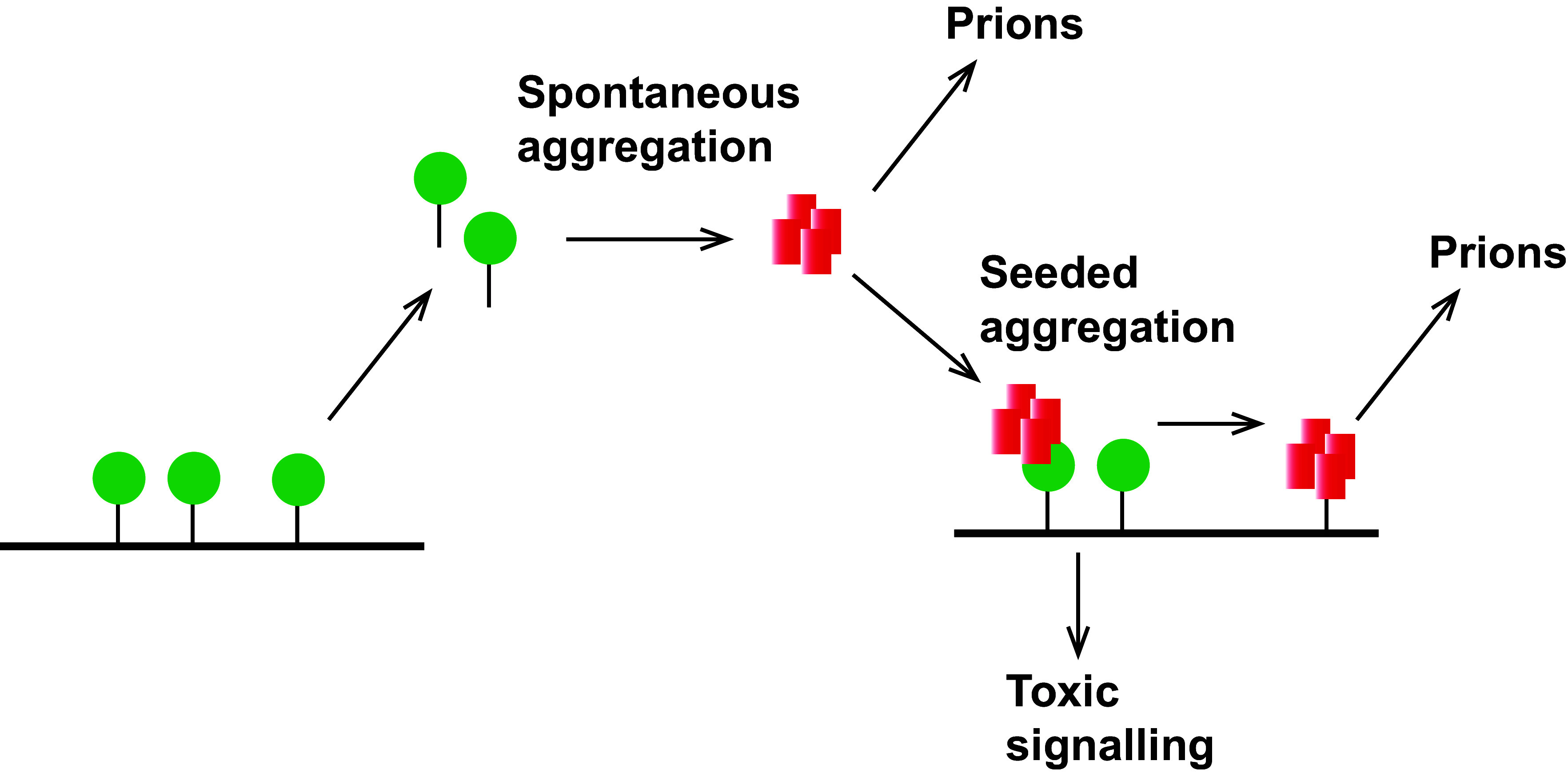
Protein misfolding and aggregation are a hallmark of various neurodegenerative disorders. However, the underlying mechanisms driving protein misfolding in the cellular context are incompletely understood. Here, we show that the two-dimensional confinement imposed by a membrane anchor stabilizes the native protein conformation and suppresses liquid-liquid phase separation (LLPS) and protein aggregation. Inherited prion diseases in humans and neurodegeneration in transgenic mice are linked to the expression of anchorless prion protein (PrP), suggesting that the C-terminal glycosylphosphatidylinositol (GPI) anchor of native PrP impedes spontaneous formation of neurotoxic and infectious PrP species. Combining unique in vitro and in vivo approaches, we demonstrate that anchoring to membranes prevents LLPS and spontaneous aggregation of PrP. Upon release from the membrane, PrP undergoes a conformational transition to detergent-insoluble aggregates. Our study demonstrates an essential role of the GPI anchor in preventing spontaneous misfolding of PrPC and provides a mechanistic basis for inherited prion diseases associated with anchorless PrP.

Keywords: liquid-liquid phase separation; membrane; neurodegenerative diseases; prion; protein aggregation.


Conflict of interest statement

Competing interests statement:The authors declare no competing interest.

Figures

Fig. 1.
Fig. 1.
A membrane anchor suppresses liquid–solid phase separation of PrP. (A) Schematic representation of the recombinant prion protein fusion protein (MBP–PrP–GFP and MBP–C2-PrP–GFP) anchored to a SLB. The N-terminal MBP can be cut off with TEV protease. The C-terminal His tag (6xHis) tethers the protein to the SLB via an interaction with NTA–Ni lipids. The protein can be liberated from the membrane via cleavage with 3C protease. C2-PrP: N-terminal truncated PrP fragment that starts at aa 90; β: beta-strands; α: alpha helices; S-S: disulfide bridge. (B) SLBs containing rhodamine DHPE were analyzed by laser scanning microscopy. Lipid mobility was measured by FRAP. After 10 s of baseline recording (prebleach), a small area of interest (AOI) was photobleached. The average normalized fluorescence intensity of AOI was plotted over time. (C) MBP–PrP–GFP (300 nM in 10 mM Tris pH 7.4, 150 mM NaCl) was incubated with TEV protease for 20 min and analyzed by laser scanning microscopy before and after TEV protease treatment (scale bar, 10 µm). (D) MBP–PrP–GFP (300 nM in 10 mM Tris pH 7.4, 150 mM NaCl) was tethered to NTA–Ni lipids in the SLB. After washing to remove unbound PrP, the SLB was incubated with TEV protease for 20 min and analyzed by laser scanning microscopy before and after TEV protease treatment (scale bar, 10 µm). (E) MBP-PrP-GFP (300 nM in 10 mM Tris pH 7.4, 150 mM NaCl) was added to SLBs lacking NTA-Ni. The sample was incubated with TEV protease for 20 min and analyzed by laser scanning microscopy before and after TEV protease treatment (scale bar, 10 µm). (F) N2a cells transiently expressing PrPΔGPI and wild-type (WT) PrPC were lysed in detergent buffer and separated into a detergent-soluble (S) and -insoluble (P) fraction by centrifugation. The fractions were analyzed by Western blotting using the anti-PrP antibody 3F4.
Fig. 2.
Fig. 2.
PrP and α-synuclein seeds induce aggregation of membrane-anchored PrP. (A) Schematic outline of the experiment. MBP–PrP–GFP (300 nM in 10 mM Tris pH 7.4, 150 mM NaCl) was tethered to NTA-Ni lipids in the SLB. After washing to remove unbound PrP, MBP was cleaved with TEV protease. The membrane-bound PrP was then incubated with preformed protein seeds. (B and C) Membrane-anchored PrP–GFP was incubated with preformed recombinant PrP seeds for 20 min. (B) The samples were analyzed by laser scanning microscopy before and after incubation with seeds (scale bar, 10 µm). (C) Volumetric 3D reconstructions generated with IMARIS software of the aggregates. Red: SLB, green: PrP-GFP. (D) Membrane-anchored PrP-GFP was incubated with preformed recombinant α-Syn seeds for 20 min and analyzed by laser scanning microscopy before and after incubation with seeds (scale bar, 10 µm). (E) Membrane-anchored PrP–GFP was incubated with soluble recombinant α-Syn for 20 min and analyzed by laser scanning microscopy before and after incubation (scale bar, 10 µm). (F) Membrane-anchored PrP–GFP was incubated with soluble recombinant MBP–PrP for 20 min and analyzed by laser scanning microscopy before and after incubation (scale bar, 10 µm).
Fig. 3.
Fig. 3.
Membrane release induces the spontaneous formation of PrP aggregates. (A and B) MBP–PrP–GFP (A) or MBP–C2-PrP–GFP (B) (300 nM in 10 mM Tris pH 7.4, 150 mM NaCl) were tethered to NTA-Ni lipids in the SLB. After washing to remove unbound PrP, membrane-anchored PrP was first incubated with TEV protease to cleave MBP and then incubated with 3C protease to release PrP–GFP from the membrane. The samples were analyzed by laser scanning microscopy before and after the addition of TEV protease and after incubation with 3C protease (scale bar, 10 µm). Volumetric 3D reconstructions generated with IMARIS software of the PrP aggregates after 3C treatment are shown on the right. Red: SLB, green: PrP-GFP. (C) Using IMARIS software, the average volume of the aggregates per sample was calculated in three independent replicates. Shown is the mean ± SD. Statistical analysis: Mann–Whitney u test, **P ≤ 0.01. (D) Scheme of GPI-anchored PrP with the 3C protease cleavage in front of the GPI anchor (PrP-3C–GPI) (E) Schematic outline of the experimental approach. N2a cells transiently expressing PrP-3C–GPI (shown in green on the cell surface) were incubated with 3C protease to release PrP-3C into the media. The cell lysates and the media were fractionated into detergent-soluble (Sup) and -insoluble fractions (Pellet) and analyzed by Western blotting (Shown in D). (F) N2a cells transiently expressing PrP-3C–GPI were treated with 3C protease (3 mg/mL) or PBS (−3C) for 4 h at 37 °C. Cell lysates and media were separated into a detergent-soluble (S) and -insoluble (P) fraction and analyzed by Western blotting using an antibody against PrP. Statistical analysis: Mann–Whitney U test, one tailed, * = 0.05.
Fig. 4.
Fig. 4.
Schematic summary of the findings. The membrane anchor stabilizes the native conformation of PrPC (green). After membrane release, PrP spontaneously misfolds into seeding-competent aggregates that could interact with and induce misfolding of membrane-anchored PrPC. This may initiate the formation of infectious prions and further initiate toxic signaling by the PrPC/Seed complex at the plasma membrane.

Similar articles

References

    1. Prusiner S. B., Prions. Proc. Natl. Acad. Sci. U.S.A. 95, 13363–13383 (1998). - PMC - PubMed
    1. Mead S., Lloyd S., Collinge J., Genetic factors in mammalian prion diseases. Annu. Rev. Genet. 53, 117–147 (2019). - PubMed
    1. Jansen C., et al. , Prion protein amyloidosis with divergent phenotype associated with two novel nonsense mutations in PRNP. Acta Neuropathol. 119, 189–197 (2010). - PMC - PubMed
    1. Chesebro B., et al. , Anchorless prion protein results in infectious amyloid disease without clinical scrapie. Science 308, 1435–1439 (2005). - PubMed
    1. Stohr J., et al. , Spontaneous generation of anchorless prions in transgenic mice. Proc. Natl. Acad. Sci. U.S.A. 108, 21223–21228 (2011). - PMC - PubMed

LinkOut - more resources