. 2025 Jan 7;122(1):e2400743122.
doi: 10.1073/pnas.2400743122. Epub 2024 Dec 31.

Memory control deficits in the sleep-deprived human brain

Affiliations

Memory control deficits in the sleep-deprived human brain

Marcus O Harrington et al. Proc Natl Acad Sci U S A. .

Abstract

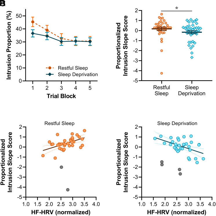
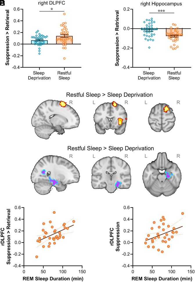
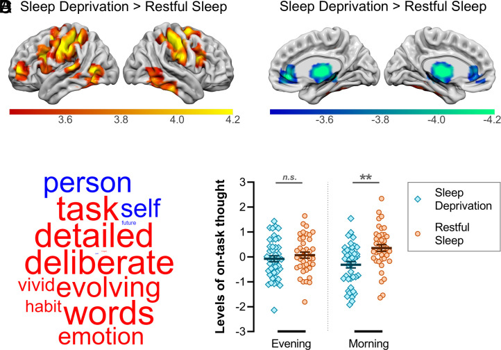
Sleep disturbances are associated with intrusive memories, but the neurocognitive mechanisms underpinning this relationship are poorly understood. Here, we show that sleep deprivation disrupts prefrontal inhibition of memory retrieval, and that the overnight restoration of this inhibitory mechanism is associated with time spent in rapid eye movement (REM) sleep. The functional impairments arising from sleep deprivation are linked to a behavioral deficit in the ability to downregulate unwanted memories, and coincide with a deterioration of deliberate patterns of self-generated thought. We conclude that sleep deprivation gives rise to intrusive memories via the disruption of neural circuits governing mnemonic inhibitory control, which may rely on REM sleep.

Keywords: default mode network; heart rate variability; inhibitory control; memory suppression; sleep deprivation.


Conflict of interest statement

Competing interests statement:The authors declare no competing interest.

Figures

Fig. 1.
Fig. 1.
Experimental procedure. (A) Timeline. The evening session began with a resting electrocardiography (ECG) recording. Participants then completed the first of two multidimensional experience sampling (MDES) tasks and the TNT learning phase, before either sleeping overnight in the laboratory where their sleep was recorded with PSG (restful sleep group) or remaining awake throughout the night (sleep deprivation group). The morning session began with another resting ECG recording. Participants then completed the TNT assessment phase inside a MRI scanner, after which a RS scan was acquired. Finally, participants completed the MDES task again. (B) MDES task. Participants monitored pairs of shapes. After 2 to 5 of these nontarget trials, a target trial occurred, wherein an additional shape appeared in the center of the screen, prompting participants to press a button corresponding to the side of the screen that the matching shape appeared on the present trial (0-back) or the immediately preceding trial (1-back). Occasionally, instead of a target trial, participants were required to indicate via a rating scale (1 to 10) the extent to which the contents of their ongoing thoughts matched a series of 13 thought probes. (C) TNT task. In the TNT learning phase, participants memorized 48 face-scene pairs. In the TNT assessment phase, faces were shown in isolation inside red or green frames. For red-framed faces (No-Think trials), participants were instructed to suppress (i.e., avoid thinking about) the associated scene. For green-framed faces (Think trials), participants were instructed to visualize the associated scene. After each trial, participants reported the extent to which they thought about the paired scene (never, briefly, or often).
Fig. 2.
Fig. 2.
Adaptive memory suppression. (A) Intrusions decreased across trial blocks more rapidly after restful sleep as compared to sleep deprivation. (B) Intrusion slope scores were lower after sleep deprivation than restful sleep (higher intrusion slope scores indicate a greater reduction in intrusions over trials). (C) In the restful sleep group, HF-HRV was positively correlated with intrusion slope scores. (D) In the sleep deprivation group, HF-HRV was negatively correlated with intrusion slope scores. *P < 0.05.
Fig. 3.
Fig. 3.
Functional brain responses during memory suppression. ROI analyses: (A) reduced engagement of the rDLPFC and (B) weaker disengagement of the right hippocampus after sleep deprivation relative to restful sleep, demonstrated by between-group differences in suppression > retrieval contrasts. Exploratory whole-brain analyses: (C) increased activation in the right superior frontal gyrus and right insular cortex, and (D) decreased activation in the right hippocampus after restful sleep (contrasted with sleep deprivation). (E) REM sleep duration was correlated with suppression-related activity in rDLPFC in the restful sleep group (suppression > retrieval contrast). (F) REM sleep duration was correlated with rDLPFC activation during suppression (as compared to baseline) in the restful sleep group, whereas no such effect was observed for activation during retrieval. *P < 0.05; ***P < 0.001, based on independent t tests. R = right hemisphere; L = left hemisphere.
Fig. 4.
Fig. 4.
RS functional connectivity and self-generated patterns of thought. (A) Sleep deprivation (in contrast to restful sleep) increased functional connectivity between the DMN and several areas of the CCN. (B) Sleep deprivation (in contrast to restful sleep) decreased functional connectivity between the DMN and thalamus bilaterally. (C) The application of principal components analysis (PCA) to MDES data identifies latent patterns of thought by grouping thought probes that capture shared variance. The first component identified from the PCA corresponded to a pattern of on-task thinking. The loadings on this component are presented as a word cloud. The color of a word describes the direction of the relationship (red: positive, blue: negative) and the size of a word reflects the magnitude of the loading. (D) Patterns of on-task thinking were significantly reduced after sleep deprivation relative to restful sleep (no such difference emerged in the prior evening session). **P < 0.01; n.s. = nonsignificant.

Similar articles

Cited by

References

    1. Brewin C. R., Gregory J. D., Lipton M., Burgess N., Intrusive images in psychological disorders: Characteristics, neural mechanisms, and treatment implications. Psychol. Rev. 117, 210–232 (2010). - PMC - PubMed
    1. Benoit R. G., Hulbert J. C., Huddleston E., Anderson M. C., Adaptive top-down suppression of hippocampal activity and the purging of intrusive memories from consciousness. J. Cogn. Neurosci. 27, 96–111 (2015). - PubMed
    1. Gagnepain P., Hulbert J., Anderson M. C., Parallel regulation of memory and emotion supports the suppression of intrusive memories. J. Neurosci. 37, 6423–6441 (2017). - PMC - PubMed
    1. Hellerstedt R., Johansson M., Anderson M. C., Tracking the intrusion of unwanted memories into awareness with event-related potentials. Neuropsychologia 89, 510–523 (2016). - PMC - PubMed
    1. Levy B. J., Anderson M. C., Purging of memories from conscious awareness tracked in the human brain. J. Neurosci. 32, 16785–16794 (2012). - PMC - PubMed

LinkOut - more resources