. 2025 Jan 7;122(1):e2419610121.
doi: 10.1073/pnas.2419610121. Epub 2024 Dec 31.

In situ architecture of a nucleoid-associated biomolecular co-condensate that regulates bacterial cell division

Affiliations

In situ architecture of a nucleoid-associated biomolecular co-condensate that regulates bacterial cell division

Peng Xu et al. Proc Natl Acad Sci U S A. .

Abstract

In most bacteria, cell division depends on the tubulin-homolog FtsZ that polymerizes in a GTP-dependent manner to form the cytokinetic Z-ring at the future division site. Subsequently, the Z-ring recruits, directly or indirectly, all other proteins of the divisome complex that executes cytokinesis. A critical step in this process is the precise positioning of the Z-ring at the future division site. While the divisome proteins are generally conserved, the regulatory systems that position the Z-ring are more diverse. However, these systems have in common that they modulate FtsZ polymerization. In Myxococcus, PomX, PomY, and PomZ form precisely one MDa-sized, nonstoichiometric, nucleoid-associated assembly that spatiotemporally guides Z-ring formation. Here, using cryo-correlative light and electron microscopy together with in situ cryoelectron tomography, we determine the PomXYZ assembly's architecture at close-to-live conditions. PomX forms a porous meshwork of randomly intertwined filaments. Templated by this meshwork, the phase-separating PomY protein forms a biomolecular condensate that compacts and bends the PomX filaments, resulting in the formation of a selective PomXYZ co-condensate that is associated to the nucleoid by PomZ. These studies reveal a hitherto undescribed supramolecular structure and provide a framework for understanding how a nonstoichiometric co-condensate forms, maintains number control, and nucleates GTP-dependent FtsZ polymerization to precisely regulate cell division.

Keywords: bacterial cell division; biomolecular condensate; correlative cryo-electron tomography; cryo-electron microscopy; liquid-liquid phase separation.


Conflict of interest statement

Competing interests statement:The authors declare no competing interest.

Figures

Fig. 1.
Fig. 1.
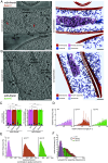
Identification of the PomXYZ assembly by CLEM and cryo-ET. (A) Correlation of mCh-PomX signal and Hoechst signal with cryo-ET square overview map at low magnification. White arrows indicate lamellae. (Scale bar, 20 µm.) (B) Correlation of mCh-PomX signal and Hoechst signal with cryo-ET lamella map at higher magnification. White arrows indicate target cells on lamella. (Scale bar, 2 μm.) (C) Correlation of mCh-PomX signal with a reconstructed tomographic slice. The black arrow indicates elongated filament bundles, white arrows mCh-PomX signals from outside the focal plane of cells removed by FIB milling, and red arrow mCh-PomX target signal. (Scale bar 100 nm.) (D and E). Left: Single image slice of a denoised tomogram of cells expressing mCh-PomX (D) or PomY-mCh (E). (Scale bar, 100 nm.) Right: The same images as on the Left; the nucleoid area is indicated in light blue and the PomXYZ assembly area in light purple, outer membrane (OM), inner membrane (IM), and ribosome (RB).
Fig. 2.
Fig. 2.
The PomXYZ co-condensate is selective and excludes ribosomes. (A) 3D rendering of features from the tomogram of Fig. 1D (Upper part) expressing mCh-PomX. Lower part, zoomed-in view of a boxed region from the Upper part. (B) 3D plot of individual nucleoid filament fragments and PomX filament fragments in the PomXYZ co-condensate. Arrows indicate orientation of individual filaments. (C) Histogram of the angles between individual nucleoid and PomX filament fragments to their corresponding averaged orientation. (D) Distribution of distances between nucleoid and PomXYZ co-condensate.
Fig. 3.
Fig. 3.
Function of PomY and PomZ in the PomXYZ co-condensate. (A and B) Tomographic slice of a ΔpomY (A) and ΔpomZ (C) cell (Left) and relative 3D segmentations of the tomograms (Right). Abbreviations and black arrows as in Fig. 1D. Red arrows point to larger protein densities inside the PomXZ assembly. (Scale bar, 100 nm.) (C) Quantification of the long (L) and short (S) axes of mCh-PomX complexes based on SIM images (Left) and Pom assemblies based in tomograms (Right). Number of cells and tomograms analyzed (n) is indicated. Colored triangles indicate the mean from three biological replicates and black line the median. Not significant (NS) P > 0.05, ** P ≤ 0.01, *** P ≤ 0.0001 based on ANOVA one-way test. (D) Histogram of the relative angle between individual Pom filament fragment orientation against the average filament orientation in strains of the indicated genotypes. (E) Histogram of the distances between Pom coordinates to its assembly spatial center. (F). Distribution of distances between nucleoid and Pom assembly in cells of indicated genotypes. *** P ≤ 0.0001 based on ANOVA one-way test.
Fig. 4.
Fig. 4.
A repeating tetrameric unit forms the PomX filaments. (A) Upper part: AlphaFold-Multimer prediction of PomX monomer structure; model is colored by the predicted Local Distance Difference Test (pLDDT) score. Scores are indicated by blue to red color gradient. The N and C termini are indicated. Lower part: Schematic of the sequence and structure features of PomX based on AlphaFold-Multimer prediction. (B) Proposed structural model of PomX tetramer based on AlphaFold-Multimer prediction, colored by chain. The N and C termini are indicated. (C) Docking of proposed structural model of PomX tetramer into the cryo-EM SPA density map of PomX filament. Colors as in B. (D) Model of PomX self-assembly and elongation. Black arrows indicate filament’s directions. (E) In vitro pull-down experiments with purified PomX214-405-Strep and PomX1-213-His6. Instant Blue-stained SDS-PAGE shows load (L), flow-through (F), wash (W), and elution (E) fractions using Strep-Tactin beads in pull-down experiments with 10 µM of the indicated proteins alone or premixed as indicated on top. Molecular size markers are shown in lanes marked M. Note that PomX1-213-His6 migrates aberrantly and according to a higher molecular weight (10). All samples were analyzed on the same gel and black lines are included for clarity. (F) Integrative hypothetical model of PomXYZ co-condensate. Green arrows indicate PomXYZ co-condensate translocation across the nucleoid.

Similar articles

Cited by

  • Current practices in the study of biomolecular condensates: a community comment.
    Alberti S, Arosio P, Best RB, Boeynaems S, Cai D, Collepardo-Guevara R, Dignon GL, Dimova R, Elbaum-Garfinkle S, Fawzi NL, Fuxreiter M, Gladfelter AS, Honigmann A, Jain A, Joseph JA, Knowles TPJ, Lasker K, Lemke EA, Lindorff-Larsen K, Lipowsky R, Mittal J, Mukhopadhyay S, Myong S, Pappu RV, Rippe K, Shelkovnikova TA, Vecchiarelli AG, Wegmann S, Zhang H, Zhang M, Zubieta C, Zweckstetter M, Dormann D, Mittag T. Alberti S, et al. Nat Commun. 2025 Aug 19;16(1):7730. doi: 10.1038/s41467-025-62055-8. Nat Commun. 2025. PMID: 40830340 Free PMC article.
  • In situ cryo-electron microscopy and tomography of cellular and organismal samples.
    Majumder P, Zhang P. Majumder P, et al. Curr Opin Struct Biol. 2025 Aug;93:103076. doi: 10.1016/j.sbi.2025.103076. Epub 2025 Jun 4. Curr Opin Struct Biol. 2025. PMID: 40472449 Free PMC article. Review.

References

    1. Cameron T. A., Margolin W., Insights into the assembly and regulation of the bacterial divisome. Nat. Rev. Microbiol. 22, 33–45 (2024). - PMC - PubMed
    1. Löwe J., Amos L. A., Crystal structure of the bacterial cell-division protein FtsZ. Nature 391, 203–206 (1998). - PubMed
    1. Lutkenhaus J., The ParA/MinD family puts things in their place. Trends Microbiol. 20, 411–418 (2012). - PMC - PubMed
    1. Schumacher D., et al. , The PomXYZ proteins self-organize on the bacterial nucleoid to stimulate cell division. Dev. Cell 41, 299–314.e213 (2017). - PubMed
    1. Ramm B., et al. , Biomolecular condensate drives polymerization and bundling of the bacterial tubulin FtsZ to regulate cell division. Nat. Commun. 14, 3825 (2023). - PMC - PubMed

MeSH terms

LinkOut - more resources