. 2025 Jan 7;122(1):e2413890121.
doi: 10.1073/pnas.2413890121. Epub 2024 Dec 31.

The CYLD-PARP1 feedback loop regulates DNA damage repair and chemosensitivity in breast cancer cells

Affiliations

The CYLD-PARP1 feedback loop regulates DNA damage repair and chemosensitivity in breast cancer cells

Miaomiao Zheng et al. Proc Natl Acad Sci U S A. .

Abstract

Poly(ADP-ribose) polymerase 1 (PARP1) plays a crucial role in DNA repair and genomic stability maintenance. However, the regulatory mechanisms governing PARP1 activity, particularly through deubiquitination, remain poorly elucidated. Using a deubiquitinase (DUB) library binding screen, we identified cylindromatosis (CYLD) as a bona fide DUB for PARP1 in breast cancer cells. Mechanistically, CYLD is recruited by PARP1 to DNA lesions upon genotoxic stress, where it cleaves K63-linked polyubiquitin chains on PARP1 at residues K748, K940, and K949, resulting in compromised PARP1 activation. In a reciprocal manner, PARP1 PARylates CYLD at sites E191, E231, E259, and E509, thereby enhancing its DUB activity. Consequently, depletion of CYLD leads to increased efficiency in base excision repair and confers breast cancer cells with resistance to alkylating agents. Conversely, overexpression of CYLD enhances sensitivity to PARP inhibitors (PARPi) even in homologous recombination-proficient breast cancer cells. These findings offer unique insights into the intricate interplay between CYLD and PARP1 in DNA repair, underscoring the pivotal role of targeting this regulatory axis for breast cancer chemotherapy.

Keywords: CYLD; PARP1; PARylation; deubiquitination; drug sensitivity.


Conflict of interest statement

Competing interests statement:The authors declare no competing interest.

Figures
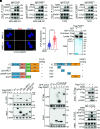
Fig. 1.
Fig. 1.
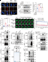
CYLD directly interacts with PARP1. (A and B) Reciprocal co-IP of endogenous CYLD and PARP1 in MDA-MB-231 cells under physiological conditions (DMSO) or DNA damage conditions (2 mM MMS, 4 h). (C and D) Reciprocal co-IP of endogenous CYLD and PARP1 in T47D cells under physiological conditions (DMSO) or DNA damage conditions (2 mM MMS, 4 h). (E) Representative images of PLA dots (red) indicating CYLD–PARP1 colocalization and DAPI (blue) in MDA-MB-231 cells under physiological conditions (DMSO) or DNA damage conditions (2 mM MMS, 2 h). (Scale bar, 10 μm.) (F) Quantification of PLA dots per cell. n ≥ 60 cells in each group were counted over three biologically independent experiments. (G) Recombinant Flag-PARP1 proteins were pulled down with either GST or GST-CYLD. Coomassie blue staining of GST or GST-CYLD protein was used as loading controls. (H and I) IP analysis to identify the domain(s) of PARP1 responsible for its interaction with CYLD achieved through cotransfection of full-length Flag-PARP1 or deletion fragments and GFP-CYLD into HEK293T cells. (J and K) IP analysis to identify the domain(s) of CYLD responsible for its interaction with PARP1 through cotransfection of full-length GFP-CYLD or deletion fragments and Flag-PARP1 into HEK293T cells. (L) IP analysis of CYLD phosphorylation in MDA-MB-231 cells with ectopically expressed Flag-CYLD using an anti-(pS/pT)Q antibody under DMSO or MMS treating conditions (2 mM, 4 h). (M) IP analysis of CYLD phosphorylation and its association with PARP1 in MDA-MB-231 cells. Cells were treated with DMSO or 2 mM MMS for 4 h before harvest.
Fig. 2.
Fig. 2.
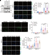
CYLD trims K63-linked polyubiquitin chains from PARP1. (A) Ubiquitination of endogenous PARP1 in MDA-MB-231 cells with ectopic expression of Myc-CYLD. (B) Ubiquitination of immunoprecipitated Flag-PARP1 by ectopically expressed GFP-CYLDWT or catalytically deficient GFP-CYLDC601S in HEK293T cells. (C) Lysates from HEK293T cells transfected with Flag-PARP1 and the combination of HA-UbK48, HA-UbK63, and GFP-CYLD were IP with anti-Flag antibody to determine which type of ubiquitin chains on PARP1 were cleaved by CYLD. (D) K63-ubiquitination of endogenous PARP1 in MDA-MB-231 cells transfected with control or CYLD siRNAs. (E) K63-ubiquitination of immunoprecipitated WT or 3 K/R mutant Flag-PARP1 in MDA-MB-231 cells transfected with control or CYLD siRNAs. 3 K/R, K748/940/949R triple mutations.
Fig. 3.
Fig. 3.
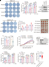
CYLD suppresses PARP1 activity in response to DNA damage. (A) Representative images of PAR (red) and DAPI (blue) staining in parental, CYLD KO, KO-WT, and KO-C/S MDA-MB-231 cells under DMSO or MMS (2 mM, 40 min) treatment conditions. (Scale bar, 25 μm.) (B) Quantification of PAR fluorescence intensity per cell. n ≥ 40 cells in each group were counted over three biologically independent experiments. (C) Representative images of XRCC1 (green) and DAPI (blue) staining in parental, CYLD KO, KO-WT, and KO-C/S MDA-MB-231 cells under DMSO or MMS (2 mM, 1 h) treatment conditions. (Scale bar, 25 μm.) (D) Quantification of XRCC1 fluorescence intensity per cell. n ≥ 60 cells in each group were counted over three biologically independent experiments. (E) Schematic of BER efficiency analysis assay. pEGFP-N1 plasmid was treated with 2 µM methylene blue in the presence of visible light (100 W high-power LED lamp) for 1 h to induce base damage. (F) Quantification of BER efficiency by flow cytometry in parental, CYLD KO, KO-WT, and KO-C/S MDA-MB-231 cells from three independent experiments. (G and H) PAR levels were detected with immunoblotting (G), and relative intensity of PAR was quantified (H) in PARP1 KD MDA-MB-231 cells stably expressing WT or 3 K/R PARP1. Cells were with or without 2 mM MMS treating for 40 min, then subjected to immunoblots with indicated antibodies.
Fig. 4.
Fig. 4.
PARP1 recruits CYLD to DNA damage sites and promotes CYLD PARylation in response to DNA damage. (A) Representative images of PLA dots (red) indicating CYLD-γH2AX colocalization and DAPI (blue) staining in MDA-MB-231 cells treated with 2 mM MMS for 2 h in the presence of PARP1 siRNA or Olaparib (5 μM, 5 h). (Scale bar, 10 μm.) (B) Quantification of PLA dots per cell. n ≥ 50 cells in each group were counted over three biologically independent experiments. (C) Chromatin-bound CYLD was detected in MDA-MB-231 cells treated with 1 mM MMS for 1 h in the presence or absence of either PARP1 siRNA or Olaparib (5 μM, 5 h). (D) Quantification of relative chromatin-bound CYLD intensity was performed across three biologically independent experiments. (E) Dynamics of GFP-tagged CYLD-NLS accumulation at DNA damage sites were detected by laser microirradiation in parental HeLa cells, with or without Olaparib treatment (5 μM, 5 h), and in PARP1 KO HeLa cells. (F) Quantification of relative GFP-CYLD-NLS fluorescence intensity at laser stripes was performed by normalizing the postdamage GFP signal to predamage (0 s) signal. Data were collected from three biologically independent experiments. (G and H) Reciprocal co-IP of endogenous CYLD and PAR in MDA-MB-231 cells. Cells were treated with 10 μM PARG inhibitor PDD00031705 for 30 min before harvest. (I) PAR levels of immunoprecipitated CYLD in T47D cells with or without PARP1 siRNA transfection. Cells were treated with 10 μM PARG inhibitor PDD00031705 for 30 min before harvest. (J) PAR levels of immunoprecipitated CYLD in MDA-MB-231 cells with or without 2 mM MMS treating for 40 min. (K) PAR levels of ectopically expressed WT or 4E/A mutant GFP-CYLD in HEK293T cells. 4E/A, E191/231/259/509A mutations. (L) PAR polymers binding capacity of purified GST, WT GST-CYLD, or 4E/A GST-CYLD mutant proteins was assessed using the dot-blot assay. (M) In vitro deubiquitylating assay by incubating immunoprecipitated Flag-PARP1 conjugated with K63-ubiquitin with PARylated WT or 4E/A mutant Myc-CYLD. The WT or 4E/A mutant Myc-CYLD was immunoprecipitated from HEK293T cells treated with 2 mM MMS for 40 min before harvest, followed by elution with Myc peptides.
Fig. 5.
Fig. 5.
CYLD exacerbates DNA damage through its DUB activity. (A) Expression levels of γH2AX in parental and CYLD KO MDA-MB-231 cells with or without 2 mM MMS treating for 6 h. (B) Representative images of γH2AX (green) and DAPI (blue) staining in parental, CYLD KO, KO-WT, and KO-C/S MDA-MB-231 cells under DMSO or MMS (1 mM, 4 h) treatment conditions. (Scale bar, 25 μm.) (C) Quantification of γH2AX fluorescence intensity per cell. n ≥ 80 cells in each group were counted over three biologically independent experiments. (D) Representative comet assay images of parental, CYLD KO, KO-WT, and KO-C/S MDA-MB-231 cells under DMSO or MMS (200 μM MMS, 40 min) treatment conditions. (Scale bar, 50 μm.) (E) Analysis of comet tail shown in (D) using the Comet Assay Software Project. (F) Representative images of γH2AX (green) and DAPI (blue) staining in SC, PARP1 KD, KD-WT, and KD-3 K/R MDA-MB-231 cells under DMSO or MMS (1 mM, 4 h) treatment conditions. (Scale bar, 25 μm.) (G) Quantification of γH2AX fluorescence intensity per cell. n ≥ 50 cells in each group were counted over three biologically independent experiments.
Fig. 6.
Fig. 6.
The CYLD–PARP1 axis sensitizes cells to DNA-damaging agents. (A and B) Representative images of colony formation of parental, CYLD KO, KO-WT, and KO-C/S MDA-MB-231 cells with or without MMS treatment at various concentrations for 14 d. MMS was added 48 h after seeding cells and replenished every 2 d. Quantification of colony numbers from three biologically independent experiments was shown in (B). (C) Expression of endogenous CYLD and exogenous Myc-tagged WT or 4E/A CYLD proteins in EV and CYLD OE MDA-MB-231 cells. (D and E) Representative images of colony formation of EV, CYLD-WT, and CYLD-4E/A MDA-MB-231 cells with or without Olaparib treatment at various concentrations for 14 d. Olaparib was added 48 h after seeding cells and replenished every 2 d. Quantification of colony numbers from three biologically independent experiments was shown in (E). (FH) Growth of EV, CYLD-WT, and CYLD-4E/A MDA-MB-231 cells in mice that were treated with Vehicle or Olaparib. Tumor image, weight and volume shown in FH, respectively. (I) Expression of PARP1 and CYLD in CYLD KO MDA-MB-231 cells with or without PARP1 siRNA transfection. (J) The growth inhibition rate of parental, CYLD KO, KO-siPARP1#1, and KO-siPARP1#2 MDA-MB-231 cells treated with MMS. Cell viability was measured using the MTT assay after treating drugs for 48 h. (K) The growth inhibition rate of SC, PARP1 KD, KD-WT, and KD-3 K/R MDA-MB-231 cells treated with MMS. Cell viability was measured using the MTT assay after treating drugs for 48 h. (L) Analysis of CYLD mRNA levels in human breast tumors and normal breast tissues. Data were retrieved from the TCGA database.

Similar articles

References

    1. Hanahan D., Weinberg R. A., Hallmarks of cancer: The next generation. Cell 144, 646–674 (2011). - PubMed
    1. Ray Chaudhuri A., Nussenzweig A., The multifaceted roles of PARP1 in DNA repair and chromatin remodelling. Nat. Rev. Mol. Cell Biol. 18, 610–621 (2017). - PMC - PubMed
    1. Bai P., Biology of poly(ADP-Ribose) polymerases: The factotums of cell maintenance. Mol. Cell 58, 947–958 (2015). - PubMed
    1. Fisher A. E. O., Hochegger H., Takeda S., Caldecott K. W., Poly(ADP-ribose) polymerase 1 accelerates single-strand break repair in concert with poly(ADP-ribose) glycohydrolase. Mol. Cell Biol. 27, 5597–5605 (2007). - PMC - PubMed
    1. Bryant H. E., et al. , PARP is activated at stalled forks to mediate Mre11-dependent replication restart and recombination. Embo J. 28, 2601–2615 (2009). - PMC - PubMed

MeSH terms

Substances

LinkOut - more resources