. 2024 Dec 31;19(12):e0312260.
doi: 10.1371/journal.pone.0312260. eCollection 2024.

"Energetics of the outer retina I: Estimates of nutrient exchange and ATP generation"

Affiliations

"Energetics of the outer retina I: Estimates of nutrient exchange and ATP generation"

Stella Prins et al. PLoS One. .

Abstract

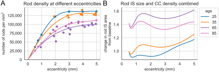
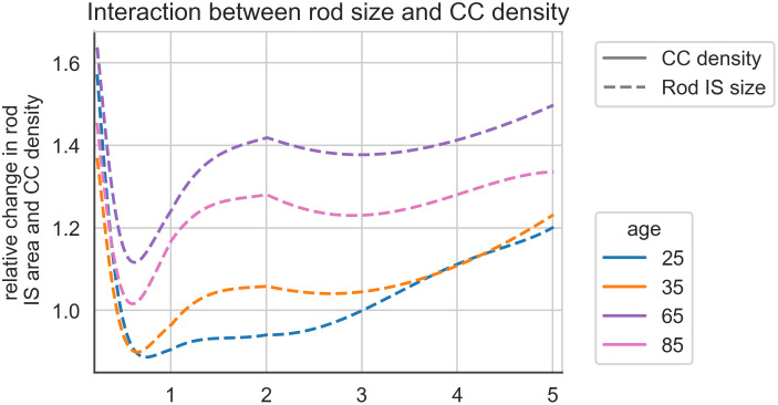
Photoreceptors (PRs) are metabolically demanding and packed at high density, which presents a challenge for nutrient exchange between the associated vascular beds and the tissue. Motivated by the ambition to understand the constraints under which PRs function, in this study we have drawn together diverse physiological and anatomical data in order to generate estimates of the rates of ATP production per mm2 of retinal surface area. With the predictions of metabolic demand in the companion paper, we seek to develop an integrated energy budget for the outer retina. It is known that rod PR number and the extent of the choriocapillaris (CC) vascular network that supports PRs both decline with age. To set the outer retina energy budget in the context of aging we demonstrate how, at different eccentricities, decline CC density is more than matched by rod loss in a way that tends to preserve nutrient exchange per rod. Together these finds provide an integrated framework for the study of outer retinal metabolism and how it might change with age.


Conflict of interest statement

The authors have declared that no competing interests exist.

Figures

Fig 1
Fig 1. Maximal ATP yield during light and dark.
The maximal ATP yield in pmol·s−1·mm−2 during light (light grey) and dark (dark grey), in absence (left panels) and presence (right panels) of amino acids for the control model and for the retina-specific models. The control model contains two coupled Human1 models, while the retina-specific models consist of an RPE model coupled to a PR rod (‘rod’) or cone (‘cone’) model.
Fig 2
Fig 2. Scatterplot depicting the inverse correlation between the percentage of flow deficits from OCTA data (Zheng et al., 2019) and CC density across different age ranges at 1 mm eccentricity from the fovea.
The fitted regression line (y = 72.6 − 1.29x) illustrates the amount at which CC density decreases as flow deficits increase. CC density was estimated using regression line coefficients of variation of CC density with age, published by Ramrattan and colleagues [10].
Fig 3
Fig 3. Retinal vascular parameters by age and eccentricity.
(A) Percentage of flow deficits in retinal vessels as detected by OCTA scans from Zheng et al., 2019, categorized by age and measured at various distances from the foveal centre. (B) Predicted CC density at different eccentricities estimated using the regression model from Fig 1. Age groups are indicated by color-coded lines and markers.
Fig 4
Fig 4. Retinal changes with age and eccentricity.
(A) Distribution of rod PR density, comparing data for four distinct age groups based on findings from Curcio et al., 1993 (specifically Figure 7A and Figure 11A). (B) Fractional changes in rod IS cross-sectional area in 4 different age groups and at varying eccentricity. Data at small eccentricities are unreliable as cones predominate and rounding errors and variability between studies become problematic.
Fig 5
Fig 5. Graph showing how fractional change in flux varies with age and eccentricity.
The solid lines give the changes predicted from altered CC density. When these changes are combined with those in rod cross sectional area (Fig 4B) the resulting shifts are shown with dotted lines.

Similar articles

Cited by

References

    1. Zouache MA, Eames I, Klettner CA, et al.. Form, shape and function: segmented blood flow in the choriocapillaris. Sci Rep 2016; 6: 35754. doi: 10.1038/srep35754 - DOI - PMC - PubMed
    1. Zouache MA, Eames I, Klettner CA, et al.. Flow and passive transport in planar multipolar flows. J Fluid Mech 2019; 858: 184–227. Journal.
    1. Zouache MA, Eames I and Luthert PJ. Blood flow in the choriocapillaris. J Fluid Mech 2015; 774: 37–66.
    1. Zouache MA. Variability in Retinal Neuron Populations and Associated Variations in Mass Transport Systems of the Retina in Health and Aging. Front Aging Neurosci 2022; 14: 778404. 20220225. doi: 10.3389/fnagi.2022.778404 - DOI - PMC - PubMed
    1. Wang L, Kondo M and Bill A. Glucose metabolism in cat outer retina. Effects of light and hyperoxia. Invest Ophthalmol Vis Sci 1997; 38: 48–55. 1997/01/01. - PubMed

Substances

LinkOut - more resources