. 2025 Feb;41(2):e12923.
doi: 10.1002/kjm2.12923. Epub 2024 Dec 30.

Curcumin-induced exosomal FTO from bone marrow stem cells alleviates sepsis-associated acute kidney injury by modulating the m6A methylation of OXSR1

Affiliations

Curcumin-induced exosomal FTO from bone marrow stem cells alleviates sepsis-associated acute kidney injury by modulating the m6A methylation of OXSR1

Ting Yang et al. Kaohsiung J Med Sci. 2025 Feb.

Abstract

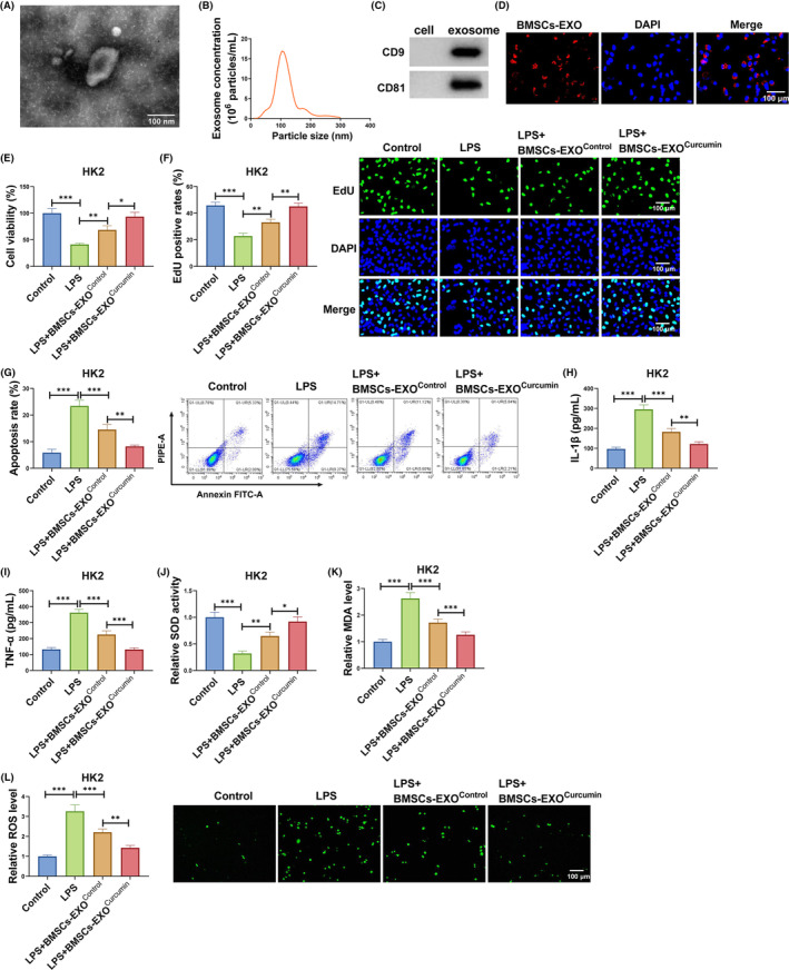
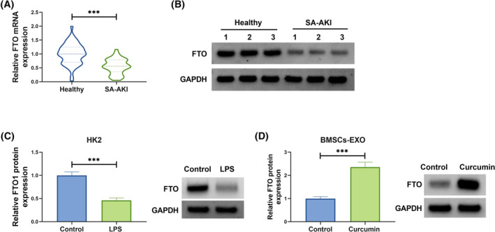
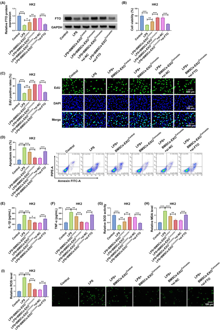
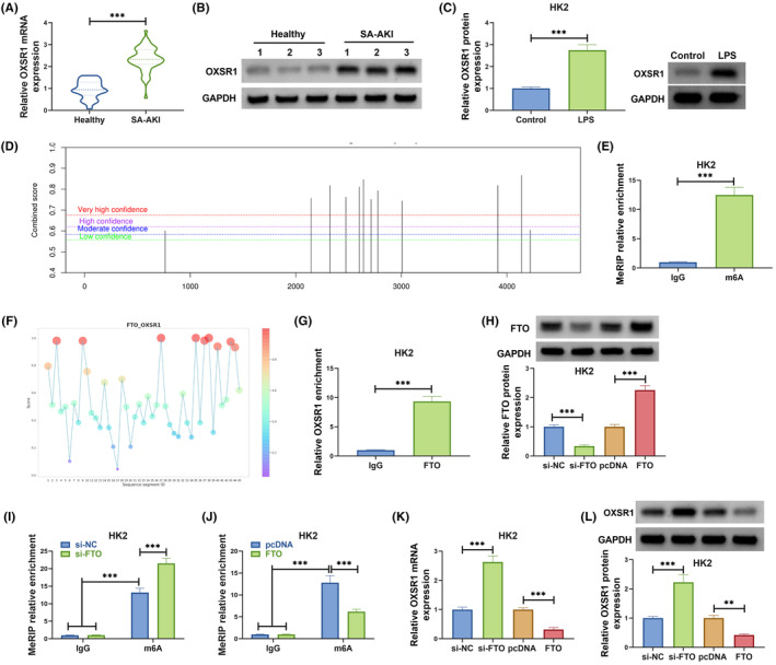
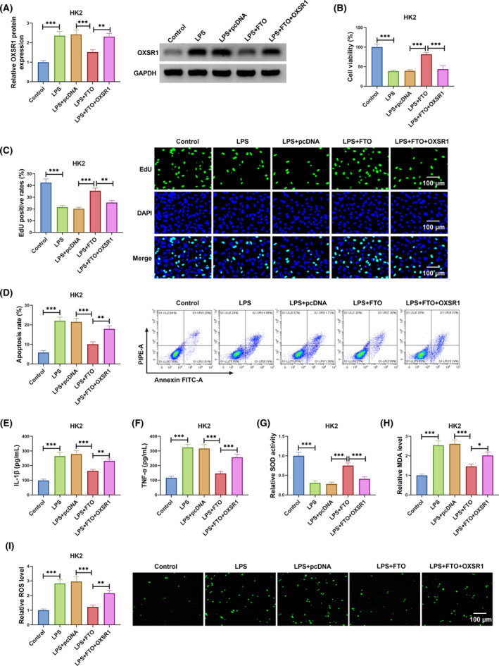
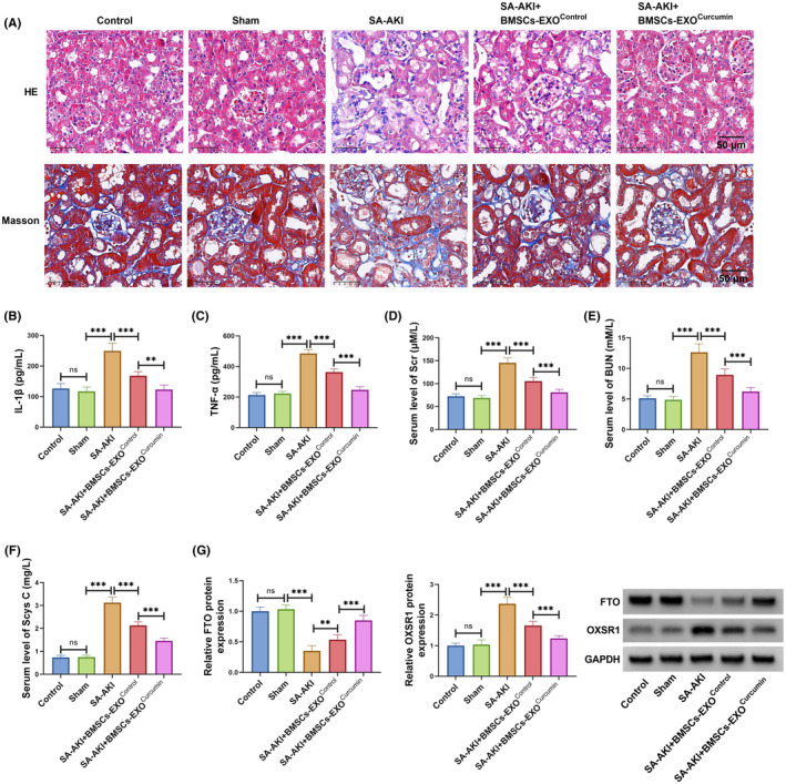
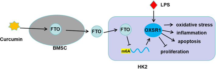
Curcumin and bone marrow stem cells (BMSCs)-derived exosomes are considered to be useful for the treatment of many human diseases, including sepsis-associated acute kidney injury (SA-AKI). However, the role and underlying molecular mechanism of curcumin-loaded BMSCs-derived exosomes in the progression of SA-AKI remain unclear. Exosomes (BMSCs-EXOCurcumin or BMSCs-EXOControl) were isolated from curcumin or DMSO-treated BMSCs, and then co-cultured with LPS-induced HK2 cells. Cell proliferation and apoptosis were determined by cell counting kit 8 (CCK8) assay, 5-ethynyl-2-deoxyuridine (EdU) assay, and flow cytometry. Enzyme-linked immunosorbent assay (ELISA) was used for examining inflammatory factors. The levels of SOD, MDA, and ROS were tested to assess oxidative stress. The levels of fat mass and obesity-associated protein (FTO) and oxidative stress responsive 1 (OXSR1) were detected by quantitative real-time PCR and western blot. Methylated RNA immunoprecipitation (MeRIP) assay and RNA immunoprecipitation (RIP) assay were used for measuring the interaction between FTO and OXSR1. BMSCs-EXOCurcumin treatment could inhibit LPS-induced HK2 cell apoptosis, inflammation, and oxidative stress. FTO was downregulated in SA-AKI patients and LPS-induced HK2 cells, while was upregulated in BMSCs-EXOCurcumin. Exosomal FTO from curcumin-induced BMSCs suppressed apoptosis, inflammation, and oxidative stress in LPS-induced HK2 cells. FTO decreased OXSR1 expression through m6A modification, and the inhibitory effect of FTO on LPS-induced HK2 cell injury could be eliminated by OXSR1 overexpression. In animal experiments, BMSCs-EXOCurcumin alleviated kidney injury in SA-AKI mice models by regulating FTO/OXSR1 axis. In conclusion, exosomal FTO from curcumin-induced BMSCs reduced OXSR1 expression to alleviate LPS-induced HK2 cell injury and improve kidney function in CLP-induced mice models, providing a new target for SA-AKI.

Keywords: FTO; OXSR1; bone marrow stem cells; exosomes; sepsis‐associated AKI.


Conflict of interest statement

The authors declare no conflicts of interest.

Figures

FIGURE 1
FIGURE 1
BMSCs‐EXOCurcumin regulated LPS‐induced HK2 cell injury. The exosomes from BMSCs was identified by TEM (A) and NTA (B). (C) WB was used to detect CD9 and CD81 in BMSCs and BMSCs‐EXO. (D) Exosome uptake assay was used to confirm the uptake of BMSCs‐EXO in HK2 cells. (E–L) LPS‐induced HK2 cells were co‐cultured with BMSCs‐EXOControl or BMSCs‐EXOCurcumin. CCK8 assay (E), EdU assay (F) and flow cytometry (G) were used to measure cell proliferation and apoptosis. (H,I) IL‐1β and TNF‐α levels were examined by ELISA. (J–L) SOD, MDA, and ROS levels were examined to assess oxidative stress. *p < 0.05, **p < 0.01, and ***p < 0.001.
FIGURE 2
FIGURE 2
FTO expression in SA‐AKI patients, LPS‐induced HK2 cells and BMSCs‐EXOCurcumin. (A,B) FTO mRNA and protein levels were examined by qRT‐PCR and WB in the serum of SA‐AKI patients and healthy controls. (C) FTO protein level was detected by WB in HK2 cells treated with or without LPS. (D) FTO protein expression in BMSCs‐EXOControl or BMSCs‐EXOCurcumin was tested by WB. ***p < 0.001.
FIGURE 3
FIGURE 3
Exosomal FTO from BMSCs‐EXOCurcumin regulated LPS‐induced HK2 cell injury. LPS‐induced HK2 cells were transfected with si‐NC/si‐FTO and co‐cultured with BMSCs‐EXOControl or BMSCs‐EXOCurcumin. (A) FTO protein expression was measured by WB. Cell proliferation and apoptosis were examined using CCK8 assay (B), EdU assay (C) and flow cytometry (D). (E,F) ELISA was used to determine IL‐1β and TNF‐α levels. (G–I) Cell oxidative stress was assessed to detect SOD, MDA, and ROS levels. *p < 0.05, **p < 0.01, and ***p < 0.001.
FIGURE 4
FIGURE 4
FTO regulated OXSR1 expression through m6A demethylation modification. (A,B) OXSR1 mRNA and protein levels in the serum of SA‐AKI patients and healthy controls were examined by qRT‐PCR and WB. (C) OXSR1 protein level in HK2 cells treated with or without LPS was detected by WB. (D) SRAMP website predicted the methylation modification sites of OXSR1. (E) MeRIP assay was used to detect the m6A level of OXSR1. (F) The binding sites between FTO and OXSR1 was predicted by RBPsuit website. (G) RIP assay was performed to confirm the interaction between FTO and OXSR1. (H) The transfection efficiencies of si‐FTO and FTO overexpression vector were confirmed by WB. (I,J) MeRIP assay was utilized to assess the effect of si‐FTO/FTO on the m6A level of OXSR1. (K,L) OXSR1 mRNA and protein levels were examined by qRT‐PCR and WB in HK2 cells transfected with si‐NC/si‐FTO/pcDNA/FTO. **p < 0.01 and ***p < 0.001.
FIGURE 5
FIGURE 5
FTO/OXSR1 axis regulated LPS‐induced HK2 cell injury. LPS‐induced HK2 cells were transfected with pcDNA/FTO/OXSR1. (A) OXSR1 protein expression was measured by WB. CCK8 assay (B), EdU assay (C) and flow cytometry (D) were performed to determine cell proliferation and apoptosis. (E,F) IL‐1β and TNF‐α levels were measured by ELISA. (G–I) The levels of SOD, MDA, and ROS were detected to evaluate cell oxidative stress. **p < 0.01 and ***p < 0.001.
FIGURE 6
FIGURE 6
BMSCs‐EXOCurcumin regulated kidney injury in CLP‐induced SA‐AKI mice models. CLP‐induced SA‐AKI mice models were injected with BMSCs‐EXOControl or BMSCs‐EXOCurcumin. (A) H&E and Masson staining was used to assess kidney injury. (B,C) IL‐1β and TNF‐α levels in the serum of mice were measured by ELISA. (D–F) The serum levels of Scr, BUN, and Scys C in mice were used to assess kidney function. (G) FTO and OXSR1 protein levels in kidney tissues were examined by WB. **p < 0.01 and ***p < 0.001.
FIGURE 7
FIGURE 7
Mechanistic diagram of the present study. Exosomal FTO from curcumin‐induced BMSCs to alleviate LPS‐induced HK2 cell oxidative stress, inflammation and apoptosis by reducing OXSR1 expression.

Similar articles

Cited by

References

    1. Koyner JL. Sepsis and kidney injury. Contrib Nephrol. 2021;199:56–70. - PubMed
    1. Kuwabara S, Goggins E, Okusa MD. The pathophysiology of sepsis‐associated AKI. Clin J Am Soc Nephrol. 2022;17(7):1050–1069. - PMC - PubMed
    1. Poston JT, Koyner JL. Sepsis associated acute kidney injury. BMJ. 2019;364:k4891. - PMC - PubMed
    1. Zarbock A, Koyner JL, Gomez H, Pickkers P, Forni L, Acute Disease Quality Initiative Group . Sepsis‐associated acute kidney injury‐treatment standard. Nephrol Dial Transpl. 2023;39(1):26–35. - PubMed
    1. Lu J, Hou Y, Liu SX, Jin B, Liu J, Li N, et al. Acetyl‐CoA synthetase 2 induces pyroptosis and inflammation of renal epithelial tubular cells in sepsis‐induced acute kidney injury by upregulating the KLF5/NF‐kappaB pathway. Cell Commun Signal. 2024;22(1):187. - PMC - PubMed

MeSH terms