. 2025 Feb:79:103443.
doi: 10.1016/j.redox.2024.103443. Epub 2024 Dec 9.

Adipocyte-derived small extracellular vesicles exacerbate diabetic ischemic heart injury by promoting oxidative stress and mitochondrial-mediated cardiomyocyte apoptosis

Affiliations

Adipocyte-derived small extracellular vesicles exacerbate diabetic ischemic heart injury by promoting oxidative stress and mitochondrial-mediated cardiomyocyte apoptosis

Lu Gan et al. Redox Biol. 2025 Feb.

Abstract

Background: Diabetes increases ischemic heart injury via incompletely understood mechanisms. We recently reported that diabetic adipocytes-derived small extracellular vesicles (sEV) exacerbate myocardial reperfusion (MI/R) injury by promoting cardiomyocyte apoptosis. Combining in vitro mechanistic investigation and in vivo proof-concept demonstration, we determined the underlying molecular mechanism responsible for diabetic sEV-induced cardiomyocyte apoptosis after MI/R.

Methods and results: Adult mice were fed a high-fat diet (HFD) for 12 weeks. sEV were isolated from plasma or epididymal adipose tissue. HFD significantly increased the number and size of plasma- and adipocyte-derived sEV. Intramyocardial injection of an equal number of diabetic plasma sEV in nondiabetic hearts significantly increased cardiac apoptosis and exacerbated MI/R-induced cardiac dysfunction. Diabetic plasma sEV significantly activated cardiac caspase 9 but not caspase 8, suggesting that diabetic sEV induces cardiac apoptosis via the mitochondrial pathway. These pathologic alterations were phenotyped by intramyocardial injection of sEV isolated from diabetic adipocytes or HGHL-challenged 3T3L1 adipocytes. To obtain direct evidence that diabetic sEV promotes cardiomyocyte apoptotic cell death, isolated neonatal rat ventricular cardiomyocytes (NRVMs) were treated with sEV and subjected to simulated ischemia/reperfusion (SI/R). Treatment of cardiomyocytes with sEV from diabetic plasma, diabetic adipocytes, or HGHL-challenged 3T3L1 adipocytes significantly enhanced SI/R-induced apoptosis and reduced cell viability. These pathologic effects were replicated by a miR-130b-3p (a molecule increased dramatically in diabetic sEV) mimic and blocked by a miRb-130b-3p inhibitor. Molecular studies identified PGC-1α (i.e. PGC-1α1/-a) as the direct downstream target of miR-130b-3p, whose downregulation causes mitochondrial dysfunction and apoptosis. Finally, treatment with diabetic adipocyte-derived sEV or a miR-130b-3p mimic significantly enhanced mitochondrial reactive oxygen species (ROS) production in SI/R cardiomyocytes. Conversely, treatment with a miR-130b-3p inhibitor or overexpression of PGC-1α extremely attenuated diabetic sEV-induced ROS production.

Conclusion: We obtained the first evidence that diabetic sEV promotes oxidative stress and mitochondrial-mediated cardiomyocyte apoptotic cell death, exacerbating MI/R injury. These pathological phenotypes were mediated by miR-130b-3p-induced suppression of PGC-1α expression and subsequent mitochondrial ROS production. Targeting miR-130b-3p mediated cardiomyocyte apoptosis may be a novel strategy for attenuating diabetic exacerbation of MI/R injury.

Keywords: Apoptosis; Diabetes; Extracellular vesicle; Myocardial ischemia/reperfusion injury.


Conflict of interest statement

Declaration of competing interest The authors declare that they have no known competing financial interests or personal relationships that could have appeared to influence the work reported in this paper.

Figures

Fig. 1
Fig. 1
The number and size of plasma- and adipocyte-derived sEV were increased in HFD mice. The plasma sEV and adipocyte sEV (Adipo-sEV) were purified from equal volumes of ND and HFD mouse plasma, culture media of primary adipocytes from equal weight of eWAT and tantamount number of 3T3L1 adipocytes by ultracentrifugation centrifugation methods. Then, the characteristics of sEV were detected. (A, D, G) Particle size distribution and number analysis using Nanosight tracking analysis. (B, E) The morphology was characterized by transmission electron microscope (TEM). (C, F, H) Western blot analysis of the exosome markers CD63, CD81 and Flotillin-1. The fold changes were calculated by comparing them to the ND sEV group (n ≥ 5, unpaired t-test).
Fig. 2
Fig. 2
Diabetic sEV promoted cardiomyocyte apoptosis by activating mitochondrial-mediated apoptotic pathways in no-diabetic mice with MI/R performance. The tantamount ND/HFD plasma sEV (A and B), ND/HFD Adipo-sEV (C and D), and NGNL/HGHL 3T3L1 sEV (E and F) were intramyocardially injected. Forty-eight hours after injection, the no-diabetic mice were subjected to MI/R. The 3 h after reperfusion, cardiomyocyte apoptotic pathways were determined by TUNEL staining (A, C and E), cleaved caspase-3, 8, 9 expressions using Western blot analysis (B, D and F) (n = 5, All experiment groups were compared to MI/R + PBS group via One-way ANOVA).
Fig. 2
Fig. 2
Diabetic sEV promoted cardiomyocyte apoptosis by activating mitochondrial-mediated apoptotic pathways in no-diabetic mice with MI/R performance. The tantamount ND/HFD plasma sEV (A and B), ND/HFD Adipo-sEV (C and D), and NGNL/HGHL 3T3L1 sEV (E and F) were intramyocardially injected. Forty-eight hours after injection, the no-diabetic mice were subjected to MI/R. The 3 h after reperfusion, cardiomyocyte apoptotic pathways were determined by TUNEL staining (A, C and E), cleaved caspase-3, 8, 9 expressions using Western blot analysis (B, D and F) (n = 5, All experiment groups were compared to MI/R + PBS group via One-way ANOVA).
Fig. 3
Fig. 3
Diabetic sEV promoted cardiomyocyte apoptosis by activating mitochondrial-mediated apoptotic pathways in SI/R-challenged NRVMs. The tantamount plasma sEV (A, D), Adipo-sEV (B, E), and 3T3L1 sEV (C, F) were incubated with neonatal rat ventricular myocyte (NRVM) and subsequently with simulated ischemia/reoxygenation (SI/R) administration (simulated ischemia 6 h and reoxygenation 3 h, n = 5). (A-C) Cell death was determined by LDH release (n = 5). (D-F) Cell apoptosis was determined by cleaved caspase-3, 8, 9 expressions by Western blot assay (n = 5). (All experiment groups were compared to the SI/R + PBS group via One-way ANOVA).
Fig. 4
Fig. 4
HFD adipocyte-carried miR-130b-3p exacerbated SI/R-induced mitochondrial-mediated apoptosis in NRVMs. (A) Expressions of cleaved caspase-3, 8. 9 were detected in NRVM cells transfected by mimics of miR-130b-3p or NC for 24 h and received SI/R administration. The protein levels were evaluated by Western blot assay (n = 5, One-way ANOVA). (B) Expressions of cleaved caspase-3, 8. 9 were detected in NRVM cells overexpressing miR-130b-3p inhibitor (inh) before HFD Adipo-sEV + SI/R administration. The cells were transfected with miR-130b-3p inh or NC inh 24 h before HFD Adipo-sEV treatment and then subjected to SI/R performance 24 h after HFD Adipo-sEV treatment. The protein levels were evaluated by Western blot assay (n = 5, One-way ANOVA).
Fig. 5
Fig. 5
miR-130b-3p decreased the expressions of diabetes-associated genes. (AB) miR-130b-3p decreased the series of diabetes-associated gene expressions. The RT [2] Profiler™ PCR Array Mouse Diabetes was performed to detect the transcripts levels in MI/R heart with NC mimic or miR-130b-3p mimic pre-administration. The changed genes were exhibited by volcano plot (A) and heatmap of downregulated differential genes (B). (n = 3, filtering criteria in A and B, fold change >1.5, multiple t-test in C, ∗p < 0.05). (C) Protein expressions were detected in NRVM cells transfected by mimics of miR-130b-3p or NC for 24 h and received SI/R administration (n = 5, One-way ANOVA). (D) Protein expressions were detected in NRVM cells overexpressing miR-130b-3p inhibitor (inh) before HFD Adipo-sEV + SI/R administration. The protein levels were evaluated by Western blot assay (n = 5, One-way ANOVA).
Fig. 6
Fig. 6
miR-130b-3p-mediated PGC-1α downregulation responsible for diabetic sEV-induced cardiomyocyte caspase-9 activation. (AD) The direct effects of miR-130b-3p upon mouse PGC-1α (the protein encoded by Ppargc1α) were identified by reporter gene analysis. The reported plasmids containing the gene mRNA CDS or 3′UTR regions (including binding sites) are shown in A and C (mutated seed sites were reversed in sequence, and red segments were seed sites). The miRNA mimics and reporter plasmids transfected 293T cells for 48 h. The regulatory effects were evaluated by firefly/renilla luciferase activity. Fold changes were calculated and normalized to the control vector (pGL3-Promoter) with miRNA mimic treatments (n = 10, Two-way ANOVA). (E) PGC-1α overexpression restored the cleaved caspase-9 protein levels in NRVM cells with miR-130b-3p mimic plus SI/R administrations. Twenty-four hours after pcDNA-f:PGC1 transfection, cells were transfected with NC or miR-130b-3p mimic. Cells were then subjected to SI/R 24 h after miR-130b-3p mimic transfection, and caspase-9 activation was determined by western blot assay (n = 5, One-way ANOVA). (F) PGC-1α overexpression restored the cleaved caspase-9 protein levels in NRVM cells with HFD Adipo-sEV plus SI/R administrations. Twenty-four hours after pcDNA-f:PGC1 transfection, cells were subjected to SI/R in the presence or absence of diabetic Adipo-sEV. The protein levels were evaluated by Western blot assay (n = 5, One-way ANOVA).
Fig. 7
Fig. 7
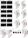
Diabetic Adiop-sEV promoted mitochondrial ROS production by miR-130b-3p-mediated PGC-1α downregulation. The mitochondria ROS production was determined by staining using the MitoSOX™ Mitochondrial Superoxide Indicators. Red fluorescence intensity indicated mitochondria ROS generation (A) miR-130b-3p promoted SI/R-induced mitochondria ROS generation in NRVMs (n = 5, One-way ANOVA). (B) miR-130b-3p inhibitors resorted HFD Adipo-sEV-induced enhancive mitochondria ROS generation in SI/R-challenged NRVMs (n ≥ 5, One-way ANOVA). (C) PGC-1α overexpression restored miR-130b-3p-induced or HFD Adipo-sEV-induced enhancive mitochondria ROS generation mimic in SI/R-challenged NRVMs (n ≥ 5, One-way ANOVA). All the intensities of red fluorescence were analyzed by Image J.
Fig. 8
Fig. 8
Diabetic adipocyte-derived sEV exacerbated cardiac injury. The tantamount ND/HFD plasma-sEV (A), ND/HFD Adipo-sEV (B), and NGNL/HGHL 3T3L1 adipocyte-derived sEV (C–E) were injected into the left anterior ventricle wall of ND mice at three different sites distal of anticipated coronary ligation. (AC) Cardiac function (LVEF and LVSF) was determined 24 h after reperfusion (n = 5, One-way ANOVA). (D) Cardiac function was evaluated by hemodynamic testing (n ≥ 5, One-way ANOVA). (E) The cardiac injury was identified by myocardial Evans blue/TTC double stain (n = 5, One-way ANOVA). (F) Schematic illustration of miR-130b-3p-enriched Adipo-sEV derived from HFD eWAT enhancing MI/R-induced mitochondrial ROS generation and mitochondrial-mediated cardiomyocyte apoptosis.
Fig. 8
Fig. 8
Diabetic adipocyte-derived sEV exacerbated cardiac injury. The tantamount ND/HFD plasma-sEV (A), ND/HFD Adipo-sEV (B), and NGNL/HGHL 3T3L1 adipocyte-derived sEV (C–E) were injected into the left anterior ventricle wall of ND mice at three different sites distal of anticipated coronary ligation. (AC) Cardiac function (LVEF and LVSF) was determined 24 h after reperfusion (n = 5, One-way ANOVA). (D) Cardiac function was evaluated by hemodynamic testing (n ≥ 5, One-way ANOVA). (E) The cardiac injury was identified by myocardial Evans blue/TTC double stain (n = 5, One-way ANOVA). (F) Schematic illustration of miR-130b-3p-enriched Adipo-sEV derived from HFD eWAT enhancing MI/R-induced mitochondrial ROS generation and mitochondrial-mediated cardiomyocyte apoptosis.

References

    1. Severino P., D'Amato A., Netti L., Pucci M., De Marchis M., Palmirotta R., Volterrani M., Mancone M., Fedele F. Diabetes mellitus and ischemic heart disease: the role of ion channels. Int. J. Mol. Sci. 2018;19 - PMC - PubMed
    1. Oikonomou E.K., Antoniades C. The role of adipose tissue in cardiovascular health and disease. Nat. Rev. Cardiol. 2018;16:83–99. - PubMed
    1. Mazzone T. Intensive glucose lowering and cardiovascular disease prevention in diabetes: reconciling the recent clinical trial data. Circulation. 2010;122:2201–2211. - PMC - PubMed
    1. Duckworth W., Abraira C., Moritz T., Reda D., Emanuele N., Reaven P.D., Zieve F.J., Marks J., Davis S.N., Hayward R., Warren S.R., Goldman S., McCarren M., Vitek M.E., Henderson W.G., Huang G.D. Glucose control and vascular complications in veterans with type 2 diabetes. N. Engl. J. Med. 2009;360:129–139. - PubMed
    1. Das Pradhan A., Glynn R.J., Fruchart J.C., MacFadyen J.G., Zaharris E.S., Everett B.M., Campbell S.E., Oshima R., Amarenco P., Blom D.J., Brinton E.A., Eckel R.H., Elam M.B., Felicio J.S., Ginsberg H.N., Goudev A., Ishibashi S., Joseph J., Kodama T., Koenig W., Leiter L.A., Lorenzatti A.J., Mankovsky B., Marx N., Nordestgaard B.G., Pall D., Ray K.K., Santos R.D., Soran H., Susekov A., Tendera M., Yokote K., Paynter N.P., Buring J.E., Libby P., Ridker P.M., Investigators P. Triglyceride lowering with pemafibrate to reduce cardiovascular risk. N. Engl. J. Med. 2022;387:1923–1934. - PubMed

Publication types

MeSH terms

LinkOut - more resources