. 2025 Jan-Feb;39(1):80-95.
doi: 10.21873/invivo.13805.

In Vivo Antitumor Activity of the PD-1/PD-L1 Inhibitor SCL-1 in Various Mouse Tumor Models

Affiliations

In Vivo Antitumor Activity of the PD-1/PD-L1 Inhibitor SCL-1 in Various Mouse Tumor Models

Tadashi Ashizawa et al. In Vivo. 2025 Jan-Feb.

Abstract

Background/aim: Immune checkpoint blockade has achieved great success as a targeted immunotherapy for solid cancers. However, small molecules that inhibit programmed death 1/programmed death ligand 1 (PD-1/PD-L1) binding are still being developed and have several advantages, such as high bioavailability. Previously, we reported a novel PD-1/PD-L1-inhibiting small compound, SCL-1, which showed potent antitumor effects on PD-L1+ tumors. These effects were dependent on CD8+ T-cell infiltration and PD-L1 expression on tumors. The present study investigated the in vivo antitumor activity of SCL-1 in various mouse syngeneic tumor models.

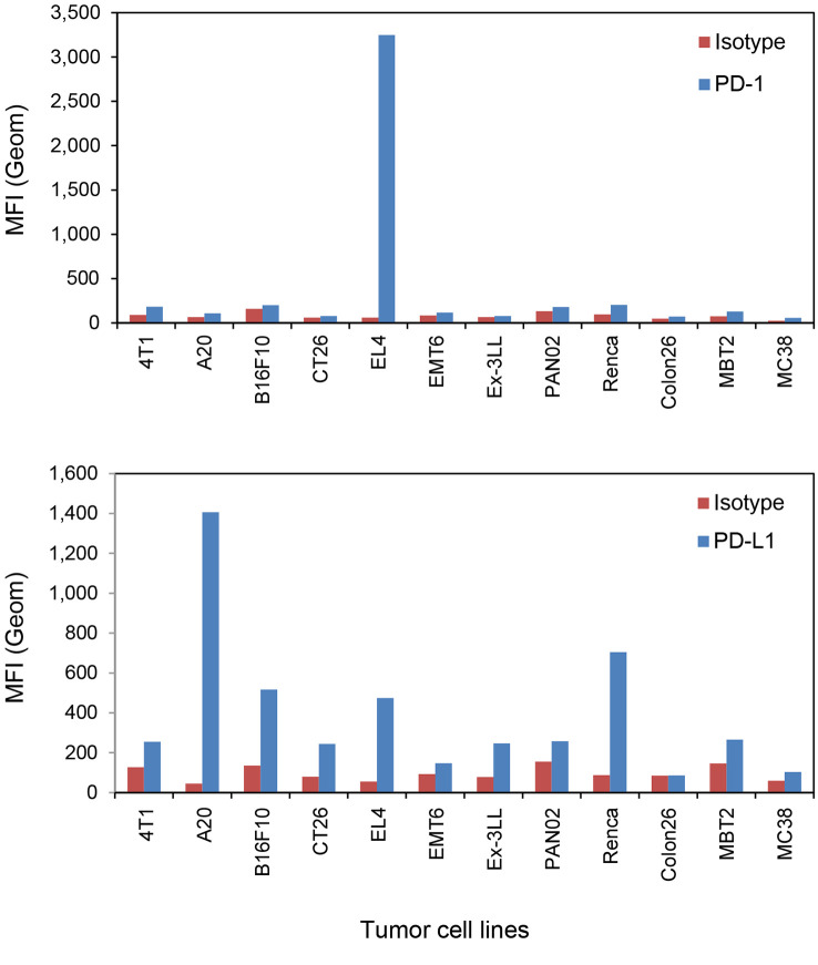
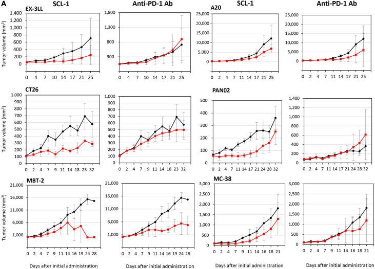
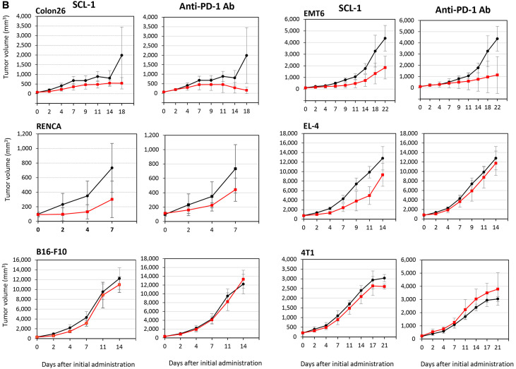
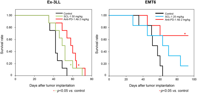
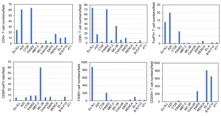
Materials and methods: Twelve syngeneic mice models of tumors, such as colon, breast, bladder, kidney, pancreatic, non-small cell lung cancers, melanoma, and lymphomas, were used for in vivo experiments. Tumor mutation burden (TMB) was analyzed by whole exome sequencing (WES) using reference DNA from mouse blood. The proportion of CD8+ T-cells infiltrating tumors before and after treatment was assessed using flow cytometry and immunohistochemistry (IHC).

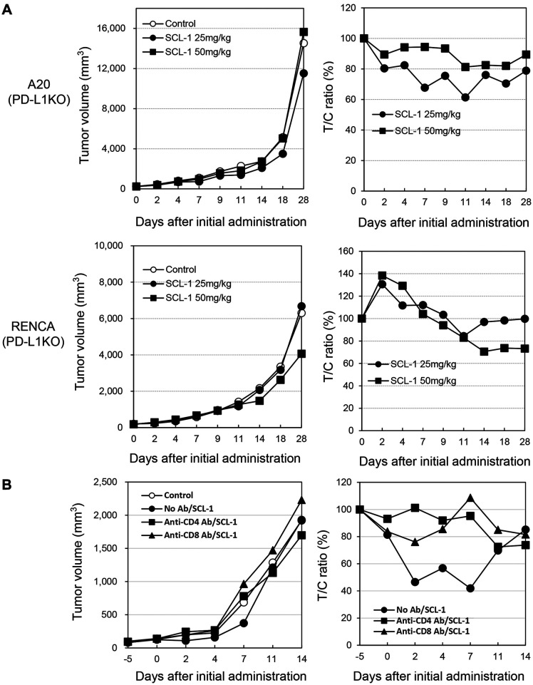
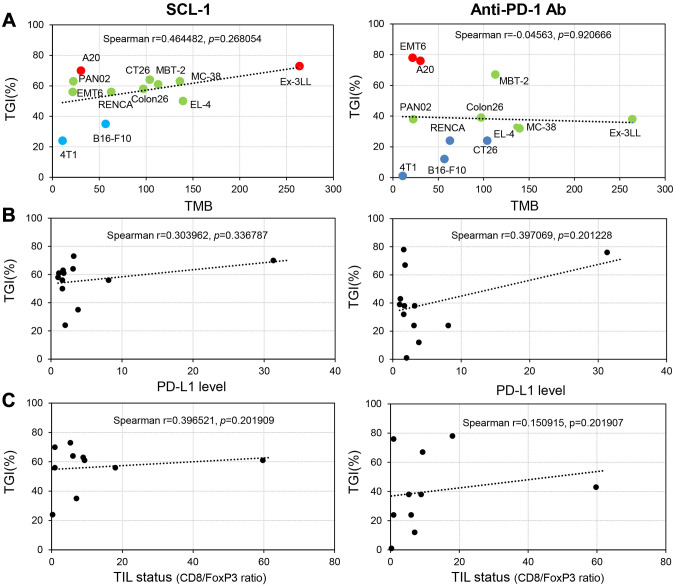
Results: SCL-1 had a markedly greater antitumor effect (11 sensitive tumors and 1 resistant tumor among the 12 tumor types) than the anti-mouse PD-1 antibody (8 sensitive tumors and 4 resistant tumors). In addition, the tumor growth inhibition rate (%) was more closely associated with TMB in the SCL-1 group than in the anti-PD-1 antibody group. Furthermore, in vivo experiments using PD-L1 gene knockout and lymphocyte-depletion technologies demonstrated that the antitumor activity of SCL-1 was dependent on CD8+ T-cell infiltration and PD-L1 expression in tumors.

Conclusion: SCL-1 has great potential as an oral immunotherapy that targets immune checkpoint molecules in cancer treatment.

Keywords: PD-1/PD-L1 inhibitor; mouse syngeneic tumors; small chemical compound; tumor mutation burden (TMB); tumor-infiltrating lymphocyte (TIL).


Conflict of interest statement

The Authors declare that they have no conflicts of interest in relation to this study.

Figures

Figure 1
Figure 1
Mouse PD-1 and PD-L1 expression levels in twelve mouse tumor cell lines determined using flow cytometry. The mean fluorescence intensity (MFI) of mouse PD-1 and PD-L1 expression is shown. In the histogram data, the MFI from the isotype antibody stain is shown in red, and the MFI from anti-mouse PD-1 or anti-mouse PD-L1 antibody stain is shown in blue. Finally, the intensity ratio (test Ab intensity/isotype Ab intensity) was calculated and used for association analysis. Each column shows the mean value from two staining experiments.
Figure 2
Figure 2
Antitumor effects of the SCL-1 compound and anti-mouse PD-1 monoclonal antibody against twelve syngeneic mouse tumors. The growth-inhibiting activity of SCL-1 or anti-PD-1 antibody is shown in Figure 2A (against Ex-3LL, CT26, MBT-2, A20, PANC02, and MC-38) and Figure 2B (against Colon26, RENCA, B16-F10, EMT6, EL-4, and 4T1). The black line represents control tumors without treatment and the red line indicates tumors treated with SCL-1 or anti-PD-1 antibody. While in most murine tumors, results at a dose of 50 mg/kg SCL-1 are shown, in RENCA tumors, results at a dose of 100 mg/kg are indicated. Anti-PD-1 Ab was administered intraperitoneally at 2 mg/kg twice weekly. Each point represents the mean±SD of six mice.
Figure 2
Figure 2
Antitumor effects of the SCL-1 compound and anti-mouse PD-1 monoclonal antibody against twelve syngeneic mouse tumors. The growth-inhibiting activity of SCL-1 or anti-PD-1 antibody is shown in Figure 2A (against Ex-3LL, CT26, MBT-2, A20, PANC02, and MC-38) and Figure 2B (against Colon26, RENCA, B16-F10, EMT6, EL-4, and 4T1). The black line represents control tumors without treatment and the red line indicates tumors treated with SCL-1 or anti-PD-1 antibody. While in most murine tumors, results at a dose of 50 mg/kg SCL-1 are shown, in RENCA tumors, results at a dose of 100 mg/kg are indicated. Anti-PD-1 Ab was administered intraperitoneally at 2 mg/kg twice weekly. Each point represents the mean±SD of six mice.
Figure 3
Figure 3
Survival benefit of the SCL-1 compound and anti-mouse PD-1 antibody in mouse syngeneic tumor models. The effect of each reagent on overall survival in tumor-bearing mice was evaluated using the Kaplan-Meier method. In the Ex-3LL tumor model (left panel), the cohorts are indicated by the black line (control group), green line (SCL-1-treated group) and red line (anti-mouse PD-1 antibody-treated). For the EMT6 tumor model (right panel), the cohorts are indicated by the black line (control group), blue line (SCL-1-treated group) and red line (anti-mouse PD-1 antibody-treated). *Statistically significant, p<0.05. Each cohort consisted of eight tumor-bearing mice.
Figure 4
Figure 4
Effect of PD-L1 gene knockout and T lymphocyte depletion on the antitumor effect of SCL-1 on mouse tumors in vivo. (A) Effect of PD-L1 gene knockout on SCL-1 antitumor activity in A20 and RENCA tumors. PD-L1 gene knockout cell clones were obtained using the CRISPR Cas9 method in clones #2 (A20) and #9 (RENCA). Tumor volumes and the ratio of tumor/control volumes are shown. (B) Impact of T-cell depletion on SCL-1 antitumor activity in RENCA tumors. Closed circle: no antibody treatment; closed square: anti-CD4 antibody treatment; closed triangle: anti-CD8 antibody treatment. *p<0.05. Each point represents the mean value of six mice.
Figure 5
Figure 5
Characterization of tumor-infiltrating immune cells before treatment using immunohistochemistry and image-analysis software. Various antibodies, such as anti-CD4, anti-CD8 and anti-FoxP3 antibodies for T-cells; anti-F4/80 antibody for macrophages; and anti-CD205 antibody for dendritic cells, were used for immune cell staining. Five fields of view in sections of each tumor at high magnification (×200) were selected, and positive cell numbers were assessed using image-analysis software. The mean positive cell count per field is shown in the blue column. Finally, the CD8+ number/FoxP3+ number ratio was calculated and used for association analysis.
Figure 6
Figure 6
Associations of in vivo tumor-growth inhibition (TGI) by SCL-1 compound or anti-PD-1 antibody with various tumor microenvironment (TME) parameters, such as the tumor mutation burden (TMB), mouse PD-L1 level and tumor-infiltrating lymphocyte (TIL) status. (A) Moderate association with TMB (r=0.464482). Mouse tumor TMB was assessed via whole-exome sequencing (WES) using tumor-derived DNA. (B) PD-L1 level and (C) TIL status were weakly associated with the TGI rate (r=0.303962 and 0.396521, respectively). TME parameter data were collected from 12 mouse tumor tissues, and the associations with the TGI rate of SCL-1 or anti-PD-1 antibody were analyzed using a Spearman coefficient test.

Similar articles

Cited by

References

    1. Topalian SL, Hodi FS, Brahmer JR, Gettinger SN, Smith DC, McDermott DF, Powderly JD, Carvajal RD, Sosman JA, Atkins MB, Leming PD, Spigel DR, Antonia SJ, Horn L, Drake CG, Pardoll DM, Chen L, Sharfman WH, Anders RA, Taube JM, McMiller TL, Xu H, Korman AJ, Jure-Kunkel M, Agrawal S, McDonald D, Kollia GD, Gupta A, Wigginton JM, Sznol M. Safety, activity, and immune correlates of anti-PD-1 antibody in cancer. N Engl J Med. 2012;366(26):2443–2454. doi: 10.1056/NEJMoa1200690. - DOI - PMC - PubMed
    1. Brahmer JR, Tykodi SS, Chow LQ, Hwu WJ, Topalian SL, Hwu P, Drake CG, Camacho LH, Kauh J, Odunsi K, Pitot HC, Hamid O, Bhatia S, Martins R, Eaton K, Chen S, Salay TM, Alaparthy S, Grosso JF, Korman AJ, Parker SM, Agrawal S, Goldberg SM, Pardoll DM, Gupta A, Wigginton JM. Safety and activity of anti-PD-L1 antibody in patients with advanced cancer. N Engl J Med. 2012;366(26):2455–2465. doi: 10.1056/NEJMoa1200694. - DOI - PMC - PubMed
    1. Wolchok JD, Kluger H, Callahan MK, Postow MA, Rizvi NA, Lesokhin AM, Segal NH, Ariyan CE, Gordon RA, Reed K, Burke MM, Caldwell A, Kronenberg SA, Agunwamba BU, Zhang X, Lowy I, Inzunza HD, Feely W, Horak CE, Hong Q, Korman AJ, Wigginton JM, Gupta A, Sznol M. Nivolumab plus ipilimumab in advanced melanoma. N Engl J Med. 2013;369(2):122–133. doi: 10.1056/NEJMoa1302369. - DOI - PMC - PubMed
    1. Sham NO, Zhao L, Zhu Z, Roy TM, Xiao H, Bai Q, Wakefield MR, Fang Y. Immunotherapy for non-small cell lung cancer: current agents and potential molecular targets. Anticancer Res. 2022;42(7):3275–3284. doi: 10.21873/anticanres.15816. - DOI - PubMed
    1. Brahmer JR, Lacchetti C, Schneider BJ, Atkins MB, Brassil KJ, Caterino JM, Chau I, Ernstoff MS, Gardner JM, Ginex P, Hallmeyer S, Holter Chakrabarty J, Leighl NB, Mammen JS, McDermott DF, Naing A, Nastoupil LJ, Phillips T, Porter LD, Puzanov I, Reichner CA, Santomasso BD, Seigel C, Spira A, Suarez-Almazor ME, Wang Y, Weber JS, Wolchok JD, Thompson JA, National Comprehensive Cancer Network Management of immune-related adverse events in patients treated with immune checkpoint inhibitor therapy: American Society of Clinical Oncology clinical practice guideline. J Clin Oncol. 2018;36(17):1714–1768. doi: 10.1200/JCO.2017.77.6385. - DOI - PMC - PubMed

MeSH terms

Substances

LinkOut - more resources