

Identification of a T2-hyperintense Perivascular Space in Brain Arteriovenous Malformations
- PMID: 39740916
- PMCID: PMC11705134
- DOI: 10.21873/invivo.13826
Identification of a T2-hyperintense Perivascular Space in Brain Arteriovenous Malformations
Abstract
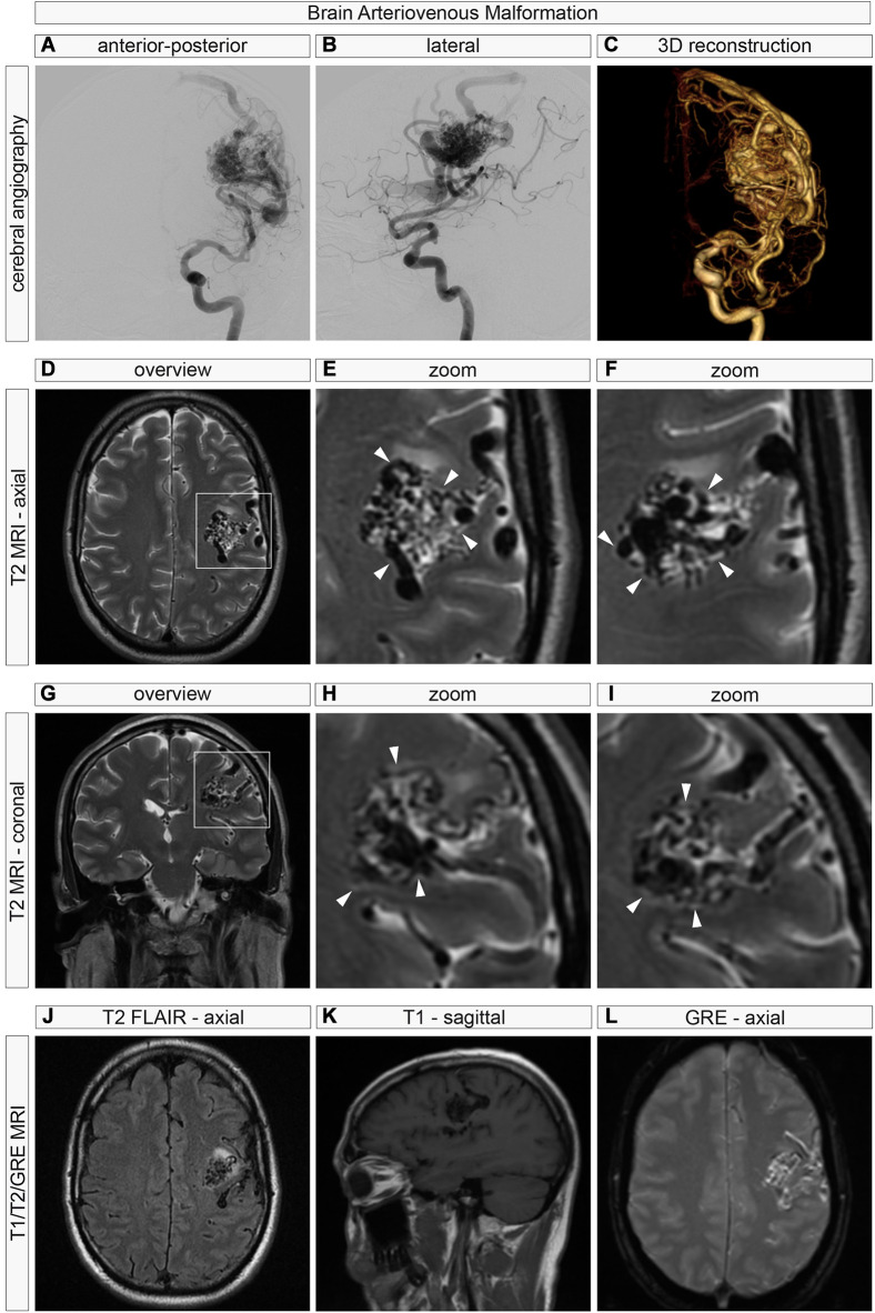
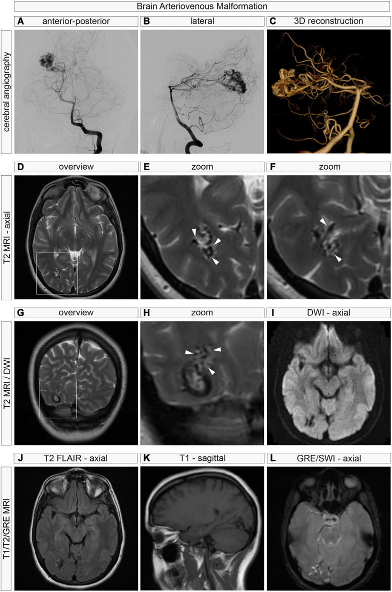
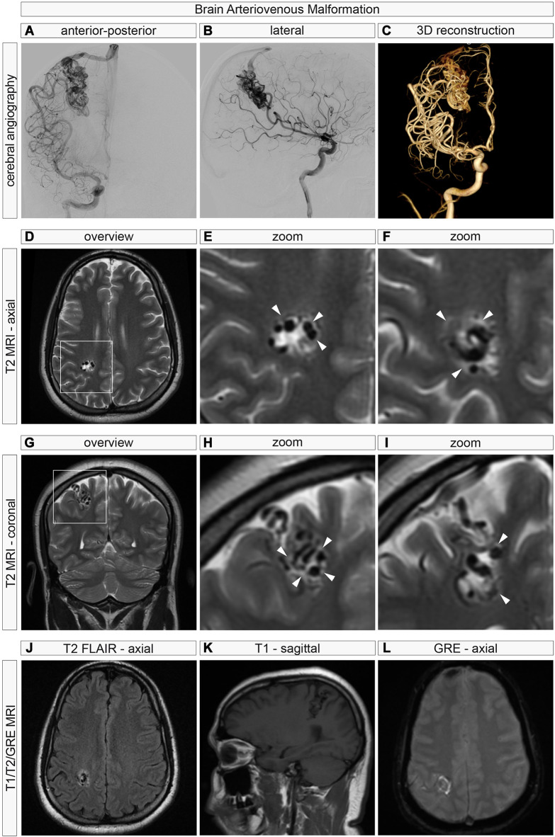
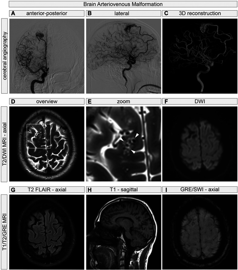
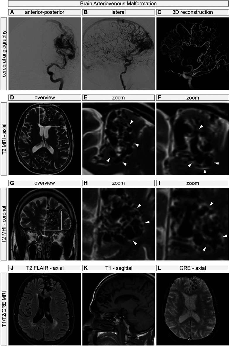
Background/aim: Brain arteriovenous malformations (AVMs) are vascular malformations characterized by dysmorphic, aberrant vasculature. During previous surgeries of compact nidus brain AVMs (representing the majority of cases), we have observed a "shiny" plane between nidal and perinidal AVM vessels and the surrounding grey and white matter and hypothesized that preoperative neuroimaging of brain AVMs may show a neuroradiological correlate of these intraoperative observations.
Patients and methods: We retrospectively reviewed and analyzed multiplanar and multisequence 3-Tesla magnetic resonance (3T MR) imaging in five consecutive brain AVMs with special attention on imaging characteristics of the brain-AVM interface, i.e., the perivascular and perinidal regions.
Results: In all five patients, we identified T2-hypertinense perivascular perinidal spaces, which were predominantly observed around the AVM nidus and less prominently around the feeding arteries or draining veins.
Conclusion: The identification of T2-hypertinense perivascular spaces surrounding brain AVMs on neuroradiological imaging may provide insights into the anatomico-radiological relationships of brain AVMs and the surrounding grey and white matter parenchyma. These findings could have future implications for our understanding of brain AVM biology and may influence neurosurgical approaches to these lesions.
Keywords: Brain AVMs; T2-hyperintense perivascular rim around AVM nidus; Virchow-Robin space; neuroradiological characterization; perivascular (perinidal) space.
Copyright © 2025, International Institute of Anticancer Research (Dr. George J. Delinasios), All rights reserved.
Conflict of interest statement
The Authors declare no competing financial interests in relation to this study.
Figures
Similar articles
-
Correlation of Appearance of MRI Perinidal T2 Hyperintensity Signal and Eventual Nidus Obliteration Following Photon Radiosurgery of Brain AVMs: Combined Results of LINAC and Gamma Knife Centers.J Neurol Surg A Cent Eur Neurosurg. 2019 May;80(3):187-197. doi: 10.1055/s-0039-1678710. Epub 2019 Mar 20. J Neurol Surg A Cent Eur Neurosurg. 2019. PMID: 30895568
-
Radiosurgery for cerebral arteriovenous malformations: assessment of early phase magnetic resonance imaging and significance of gadolinium-DTPA enhancement.Int J Radiat Oncol Biol Phys. 1996 Feb 1;34(3):663-75. doi: 10.1016/0360-3016(95)02160-4. Int J Radiat Oncol Biol Phys. 1996. PMID: 8621291 Clinical Trial.
-
Susceptibility-Based Characterization of Cerebral Arteriovenous Malformations.Invest Radiol. 2020 Nov;55(11):702-710. doi: 10.1097/RLI.0000000000000695. Invest Radiol. 2020. PMID: 32604388
-
Unruptured brain arteriovenous malformations and hydrocephalus: Case series and review of the literature.J Clin Neurosci. 2019 Jun;64:116-121. doi: 10.1016/j.jocn.2019.03.042. Epub 2019 Apr 1. J Clin Neurosci. 2019. PMID: 30948310
-
Transvenous Approach to Intracranial Arteriovenous Malformations: Challenging the Axioms of Arteriovenous Malformation Therapy?Neurosurgery. 2015 Oct;77(4):644-51; discussion 652. doi: 10.1227/NEU.0000000000000869. Neurosurgery. 2015. PMID: 26120797 Review.
Cited by
-
The Glymphatic System and Its Role in Neurovascular Diseases.J Neuroendovasc Ther. 2025;19(1):2025-0020. doi: 10.5797/jnet.ra.2025-0020. Epub 2025 Jun 21. J Neuroendovasc Ther. 2025. PMID: 40548139 Free PMC article. Review.
References
-
- Wälchli T, Ghobrial M, Schwab M, Takada S, Zhong H, Suntharalingham S, Vetiska S, Gonzalez DR, Wu R, Rehrauer H, Dinesh A, Yu K, Chen ELY, Bisschop J, Farnhammer F, Mansur A, Kalucka J, Tirosh I, Regli L, Schaller K, Frei K, Ketela T, Bernstein M, Kongkham P, Carmeliet P, Valiante T, Dirks PB, Suva ML, Zadeh G, Tabar V, Schlapbach R, Jackson HW, De Bock K, Fish JE, Monnier PP, Bader GD, Radovanovic I. Single-cell atlas of the human brain vasculature across development, adulthood and disease. Nature. 2024;632(8025):603–613. doi: 10.1038/s41586-024-07493-y. - DOI - PMC - PubMed
-
- Wälchli T, Ndengera M, Constanthin PE, Bisschop J, Morel S, Gautschi O, Berhouma M, Kalyvas A, Monnier PP, Winkler EA, Kortman H, Bhatia K, Dammann P, Jägersberg M, Gondar R, Schaller K, Kwak BR, Bijlenga P. Sex‐dependent manifestations of intracranial aneurysms. Stroke Vasc Interv Neurol. 2024;4(4):e001091. doi: 10.1161/SVIN.123.001091. - DOI
-
- Nikolaev SI, Vetiska S, Bonilla X, Boudreau E, Jauhiainen S, Rezai Jahromi B, Khyzha N, DiStefano PV, Suutarinen S, Kiehl TR, Mendes Pereira V, Herman AM, Krings T, Andrade-Barazarte H, Tung T, Valiante T, Zadeh G, Tymianski M, Rauramaa T, Ylä-Herttuala S, Wythe JD, Antonarakis SE, Frösen J, Fish JE, Radovanovic I. Somatic activating KRAS mutations in arteriovenous malformations of the brain. N Engl J Med. 2018;378(3):250–261. doi: 10.1056/NEJMoa1709449. - DOI - PMC - PubMed
MeSH terms
LinkOut - more resources
Full Text Sources
Medical