. 2025 Mar;134(3):759-771.
doi: 10.1016/j.bja.2024.09.031. Epub 2024 Dec 30.

Kappa opioid receptor internalisation-induced p38 nuclear translocation suppresses glioma progression

Affiliations

Kappa opioid receptor internalisation-induced p38 nuclear translocation suppresses glioma progression

Yong Li et al. Br J Anaesth. 2025 Mar.

Abstract

Background: Recent studies have implicated a role for perioperative medications in determining patient outcomes after surgery for malignant tumours, including relapse and metastasis.

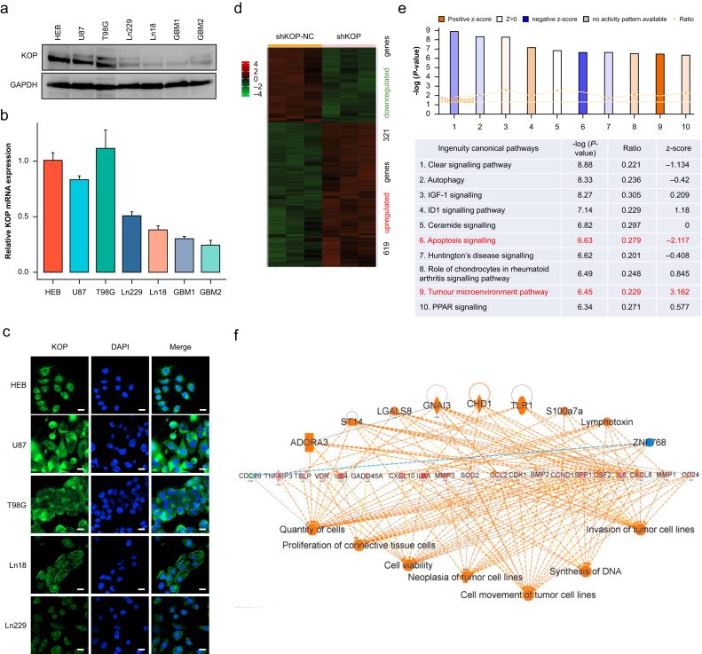
Methods: A combined approach spanned molecular, cellular, and organismal levels, including bioinformatics, immunohistochemical staining of clinical and animal samples, RNA sequencing of glioblastoma multiforme (GBM) cells with Ingenuity Pathway Analysis, lentiviral-mediated gene expression modulation, in vitro cell experiments, and in vivo orthotopic tumour transplantation.

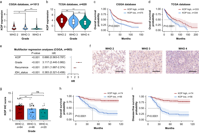
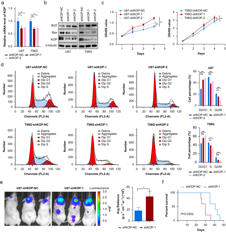
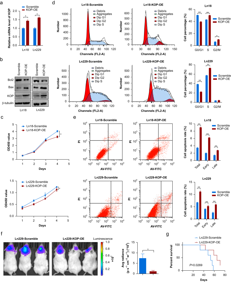
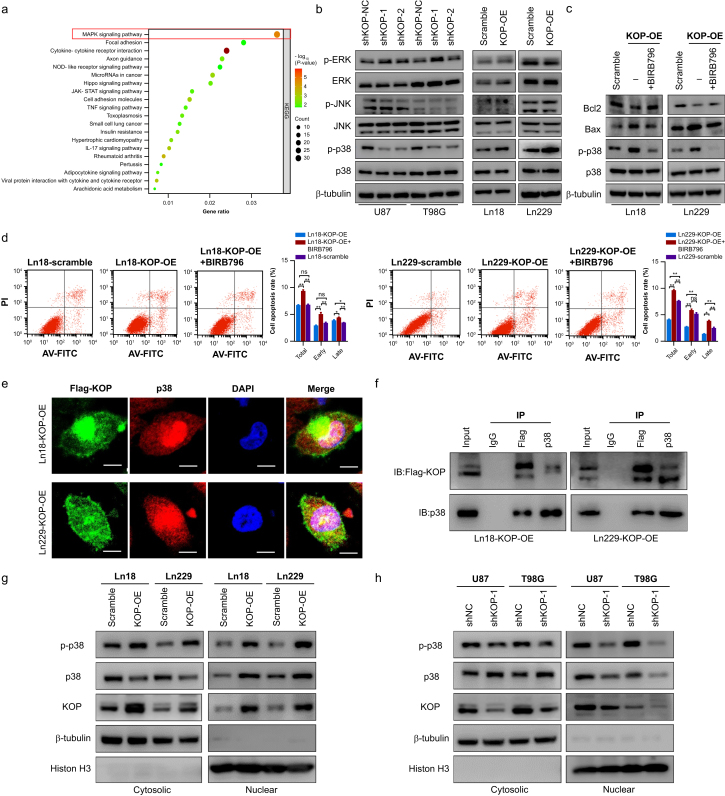
Results: We observed a significant correlation between increased kappa opioid receptor (KOP receptor) expression and better prognosis in patients with glioma. Exogenous KOP receptor overexpression in GBM cells in vitro induced cell cycle arrest, suppressed cell growth, and promoted apoptosis. Conversely, reducing KOP receptor expression in GBM cells reduced the proportion of cells in S and G2/M phases, accelerating cell growth. KOP receptor overexpression inhibited glioma cell growth and prolonged survival in mice in vivo, while KOP receptor knockdown had the opposite effect. Mechanistically, internalised KOP receptors were found to bind cytoplasmic p38, facilitating its nuclear translocation and phosphorylation, which influences downstream gene expression. The selective KOP receptor agonist TRK-820 triggered KOP receptor internalisation, activated the p38 pathway, and diminished glioma cell viability in vitro.

Conclusions: This combined molecular, cellular, and in vivo approach supports use of KOP receptor agonists as potential adjuvant therapeutics for glioma.

Keywords: KOP agonist; KOP receptor; glioblastoma multiforme; p38 MAP kinase; receptor internalisation.


Conflict of interest statement

Declaration of interest The authors declare that they have no conflicts of interest.

Figures

Fig 1
Fig 1
KOP receptor expression correlates with tumour grade and predicts better prognosis in patients with glioma. (a,b) KOP receptor expression decreases with increasing tumour grade in patients with glioma, as observed in both the CGGA (a) and TCGA (b) databases. (c,d) Kaplan–Meier survival analysis reveals that high KOP receptor expression is associated with a favourable prognosis in patients with glioma from both CGGA (c) and TCGA (d) databases. (e) Cox multifactor regression analysis utilising CGGA data indicate KOP receptor as an independent prognostic factor for patients with glioma. (f) Representative immunohistochemistry (IHC) staining images depict KOP receptor expression in clinical glioma tissue. Scale bar=100 μm. (g) IHC scores for KOP receptor are lower in high-grade glioma compared with low-grade glioma in clinical specimens. (h) And (i) Kaplan–Meier survival analysis on clinical tissue data reveals a positive correlation between high KOP receptor expression and improved overall survival (OS) (h) and disease-free survival (DFS) (i) in patients with glioma. HR, hazard ratio; IDH, isocitrate dehydrogenase; IHC, immunohistochemistry; KOP, kappa opioid; ns, not significant. ∗P<0.05. ∗∗P<0.01.
Fig 2
Fig 2
The expression pattern and functional role analysis of KOP receptor in GBM cells. (a,b) KOP receptor expression in HEB and GBM cells analysed by western immunoblotting (a) and qPCR (b). (c) Immunofluorescence detection of KOP receptor expression in HEB and GBM cells. Scale bar=20 μm. (d) RNA sequencing cluster analysis comparing KOP receptor knockdown group and control group in T98G cells. (e) Ingenuity Canonical Pathways analysis of RNA sequencing results. Red font highlights pathways with |z score|>2. (f) Results of regulatory effect analysis based on Ingenuity Pathway Analysis. GADPH, glyceraldehyde-3-phosphate dehydrogenase; GBM, glioblastoma multiforme; ID1, inhibitor of differentiation 1; IGF-1,insulin-like growth factor 1; KOP, kappa opioid; mRNA, messenger RNA; PPAR, peroxisome proliferator-activated receptor; qPCR, quantitative polymerase chain reaction.
Fig 3
Fig 3
KOP receptor knockdown promotes GBM cell survival in vitro and in vivo. (a) qPCR analysis revealed a significant decrease in KOP receptor mRNA levels in the knockdown group. (b) Western immunoblot analysis indicated significant decreases in KOP receptor and Bax proteins, along with increased Bcl2 expression, in the KOP receptor knockdown group. (c) CCK8 assay showed accelerated cell proliferation in the KOP receptor knockdown group compared with the control group. (d) Flow cytometry analysis revealed a significant increase in the proportion of cells in G0/G1 phase and a decrease in S and G2/M phase cells in the KOP receptor knockdown group compared with the control group, n=3. (e) Bioluminescence images and quantification of xenografts derived from U87 cells expressing control shRNA or shKOP-1. Images were captured on the 21st day. Data are presented as mean (standard deviation), n=5. (f) Kaplan–Meier survival analysis of tumour-bearing mice indicated a significantly shortened survival time in the KOP receptor knockdown group compared with controls. GBM, glioblastoma multiforme; KOP, kappa opioid; mRNA, messenger RNA; qPCR, quantitative polymerase chain reaction; shRNA, short hairpin RNA. ∗P<0.05. ∗∗P<0.01.
Fig 4
Fig 4
KOP receptor overexpression promotes GBM cell apoptosis and inhibits cell growth. (a) qPCR analysis revealed higher KOP receptor mRNA expression levels in the overexpression group compared with the control group. (b) Western immunoblot analysis showed an increase in KOP receptor and Bax protein expression, along with a decrease in Bcl2 expression, compared with the control group. (c) CCK8 assay demonstrated a significant reduction in cell proliferation capacity after overexpression of KOP receptor, n=3. (d) Flow cytometry analysis indicated a significant decrease in the proportion of cells in G0/G1 phase and an increase in cells in S and G2/M phases in the KOP receptor overexpression group compared with the control group, n=3. (e) Flow cytometry revealed an increase in apoptosis in the KOP receptor overexpression group compared with controls, n=3. (f) Bioluminescence images and quantification of xenografts derived from Ln229 cells expressing KOP receptor or control. Images were captured on the 21st day. Data are presented as mean (standard deviation), n=5. (g) Kaplan–Meier survival analysis of tumour-bearing mice indicated a prolonged survival time in the KOP overexpression group compared with controls. AV-FITC, annexin V-FITC; GBM, glioblastoma multiforme; KOP, kappa opioid; mRNA, messenger RNA; PI, propidium iodide; qPCR, quantitative polymerase chain reaction. ∗P<0.05. ∗∗P<0.01.
Fig 5
Fig 5
KOP receptor inhibits GBM cell survival and requires p38/MAPK pathway activation. (a) GSEA-KEGG enrichment analysis of RNA-sequencing data in the KOP receptor knockdown group revealed enrichment of the MAPK pathway. (b) western immunoblot analysis of MAPK signalling pathway-related proteins in KOP receptor knockdown and overexpression in GBM cells, and their parent cells, showed alterations in p38-MAPK expression. (c) Treatment with the p38 inhibitor BIRB796 partially reversed the KOP receptor-induced changes in apoptosis-related proteins. (d) Flow cytometry analysis demonstrated that the p38 inhibitor BIRB796 partially restored KOP receptor-induced apoptosis, n=3. (e) Immunofluorescence analysis showed KOP receptor and p38 co-localisation in both the cytoplasm and nucleus of KOP-overexpressed GBM cells. Scale bar=10 μm. (f) Immunoprecipitation analysis revealing the interaction between KOP receptor and p38. (g) After KOP receptor overexpression, KOP receptor expression increased in both the cytoplasm and nucleus, p38 distribution decreased in the cytoplasm and increased in the nucleus, while p-p38 levels increased in both the cytoplasm and nucleus. (h) After KOP receptor knockdown, KOP receptor expression decreased in both the cytoplasm and nucleus, p38 distribution increased in the cytoplasm and decreased in the nucleus, and p-p38 levels decreased in both the cytoplasm and nucleus. AV-FITC, annexin V-FITC; GBM, glioblastoma multiforme; GSEA, gene set enrichment analysis; IP, immunoprecipitation; KEGG, Kyoto Encyclopedia of Genes and Genomes; IL-17, interleukin-17; KOP, kappa opioid; MAPK, mitogen-activated protein kinase; NOD, ucleotide-binding oligomerization domain; PI, propidium iodide; TNF, tumour necrosis factor. ∗P<0.05. ∗∗P<0.01.
Fig 6
Fig 6
KOP receptor agonist TRK-820 induces KOP receptor internalisation to activate p38/MAPK signalling. (a) TRK-820 inhibits GBM cell activity in vitro at low concentrations, particularly at 5 nM, n=3. (b) Immunofluorescence reveals that TRK-820 enhanced KOP receptor nuclear translocation. (c) Western immunoblot analysis confirms that stimulation with TRK-820 5 nM markedly increases expression of KOP receptor, p38, and p-p38 proteins in the nucleus. (d) Western immunoblot analysis demonstrates that TRK-820 upregulates Bax expression and downregulates Bcl2 expression. These changes can be partially reversed by the KOP inhibitor nor-BNI and the p38 inhibitor BIRB796. (e) Treatment with Trk-820 5 nM promotes apoptosis of GBM cells, which can be largely reversed by nor-BNI and partially reversed by BIRB796, n=3. (f) TRK-820 5 nM suppresses GBM cell viability, which can be partially reversed by nor-BNI and BIRB796, n=3. (g) Schematic diagram illustrating the mechanism: KOP receptor agonists stimulate translocation of KOP receptors into the cytoplasm. Upon entering the cytoplasm, KOP receptors bind to p38 and subsequently translocate into the nucleus. Within the nucleus, p38 undergoes phosphorylation, thereby regulating expression of downstream genes. This process leads to reduced DNA synthesis, slowed cell growth, and promotion of apoptosis. AV-FITC, annexin V-FITC; DAPI, 4',6-diamidino-2-phenylindole; GBM, glioblastoma multiforme; KOP, kappa opioid; PI, propidium iodide. ∗P<0.05.

Similar articles

Cited by

References

    1. Nicholson J.G., Fine H.A. Diffuse glioma heterogeneity and its therapeutic implications. Cancer Discov. 2021;11:575–590. - PubMed
    1. Verdugo E., Puerto I., Medina M.Á. An update on the molecular biology of glioblastoma, with clinical implications and progress in its treatment. Cancer Commun (Lond) 2022;42:1083–1111. - PMC - PubMed
    1. Tan A.C., Ashley D.M., López G.Y., Malinzak M., Friedman H.S., Khasraw M. Management of glioblastoma: state of the art and future directions. CA Cancer J Clin. 2020;70:299–312. - PubMed
    1. Hiller J.G., Perry N.J., Poulogiannis G., Riedel B., Sloan E.K. Perioperative events influence cancer recurrence risk after surgery. Nat Rev Clin Oncol. 2018;15:205–218. - PubMed
    1. Casolino R., Braconi C., Malleo G., et al. Reshaping preoperative treatment of pancreatic cancer in the era of precision medicine. Ann Oncol. 2021;32:183–196. - PMC - PubMed

Substances