Optical sectioning methods in three-dimensional bioimaging
- PMID: 39741128
- PMCID: PMC11688461
- DOI: 10.1038/s41377-024-01677-x
Optical sectioning methods in three-dimensional bioimaging
Abstract
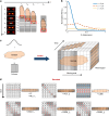
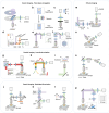
In recent advancements in life sciences, optical microscopy has played a crucial role in acquiring high-quality three-dimensional structural and functional information. However, the quality of 3D images is often compromised due to the intense scattering effect in biological tissues, compounded by several issues such as limited spatiotemporal resolution, low signal-to-noise ratio, inadequate depth of penetration, and high phototoxicity. Although various optical sectioning techniques have been developed to address these challenges, each method adheres to distinct imaging principles for specific applications. As a result, the effective selection of suitable optical sectioning techniques across diverse imaging scenarios has become crucial yet challenging. This paper comprehensively overviews existing optical sectioning techniques and selection guidance under different imaging scenarios. Specifically, we categorize the microscope design based on the spatial relationship between the illumination and detection axis, i.e., on-axis and off-axis. This classification provides a unique perspective to compare the implementation and performances of various optical sectioning approaches. Lastly, we integrate selected optical sectioning methods on a custom-built off-axis imaging system and present a unique perspective for the future development of optical sectioning techniques.
© 2025. The Author(s).
Conflict of interest statement
Conflict of interest: Q.L., J.Y., R.J., and H.G. have been licensed co-inventors of LiMo and DSIM’s patents. All authors declare that they have no other competing interests.
Figures


Similar articles
-
A phasor-based approach to improve optical sectioning in any confocal microscope with a tunable pinhole.Microsc Res Tech. 2022 Sep;85(9):3207-3216. doi: 10.1002/jemt.24178. Epub 2022 Jun 10. Microsc Res Tech. 2022. PMID: 35686877 Free PMC article.
-
DMD-based LED-illumination super-resolution and optical sectioning microscopy.Sci Rep. 2013;3:1116. doi: 10.1038/srep01116. Epub 2013 Jan 23. Sci Rep. 2013. PMID: 23346373 Free PMC article.
-
Modulated-alignment dual-axis (MAD) confocal microscopy for deep optical sectioning in tissues.Biomed Opt Express. 2014 Apr 30;5(6):1709-20. doi: 10.1364/BOE.5.001709. eCollection 2014 Jun 1. Biomed Opt Express. 2014. PMID: 24940534 Free PMC article.
-
Optical sectioning microscopy with planar or structured illumination.Nat Methods. 2011 Sep 29;8(10):811-9. doi: 10.1038/nmeth.1709. Nat Methods. 2011. PMID: 21959136 Review.
-
Grating imager systems for fluorescence optical-sectioning microscopy.Cold Spring Harb Protoc. 2014 Sep 2;2014(9):923-31. doi: 10.1101/pdb.top083493. Cold Spring Harb Protoc. 2014. PMID: 25183825 Review.
Cited by
-
Optical tweeze-sectioning microscopy for 3D imaging and manipulation of suspended cells.Sci Adv. 2025 Jul 4;11(27):eadx3900. doi: 10.1126/sciadv.adx3900. Epub 2025 Jul 2. Sci Adv. 2025. PMID: 40601731 Free PMC article.
-
Computational self-corrected quantitative 3D topographic imaging.Sci Rep. 2025 Jul 7;15(1):24298. doi: 10.1038/s41598-025-08284-9. Sci Rep. 2025. PMID: 40624205 Free PMC article.
-
Self-Supervised Z-Slice Augmentation for 3D Bio-Imaging via Knowledge Distillation.ArXiv [Preprint]. 2025 Mar 17:arXiv:2503.04843v2. ArXiv. 2025. PMID: 40093360 Free PMC article. Preprint.
-
Structured detection for simultaneous super-resolution and optical sectioning in laser scanning microscopy.Nat Photonics. 2025;19(8):888-897. doi: 10.1038/s41566-025-01695-0. Epub 2025 Jun 5. Nat Photonics. 2025. PMID: 40823346 Free PMC article.
References
-
- Pawley, J. B. Handbook of Biological Confocal Microscopy. 3rd edn. (New York: Springer, 2006).
-
- So, P. T. C. et al. Two-photon excitation fluorescence microscopy. Annu Rev. Biomed. Eng.2, 399–429 (2000). - PubMed
-
- Im, K. B. et al. Simple high-speed confocal line-scanning microscope. Opt. Express13, 5151–5156 (2005). - PubMed
-
- Tanaami, T. et al. High-speed 1-frame/ms scanning confocal microscope with a microlens and Nipkow disks. Appl Opt.41, 4704–4708 (2002). - PubMed
-
- Neil, M. A. A., Juškaitis, R. & Wilson, T. Method of obtaining optical sectioning by using structured light in a conventional microscope. Opt. Lett.22, 1905–1907 (1997). - PubMed
Publication types
Grants and funding
- 62325502/National Natural Science Foundation of China (National Science Foundation of China)
- 81827901/National Natural Science Foundation of China (National Science Foundation of China)
- 2021ZD0201001/Ministry of Science and Technology of the People's Republic of China (Chinese Ministry of Science and Technology)
- COCHE-1.5/Innovation and Technology Commission (ITF)
LinkOut - more resources
Full Text Sources
Research Materials

