Review
. 2025 Feb;57(1):1-12.
doi: 10.1038/s12276-024-01364-2. Epub 2025 Jan 1.

Trogocytosis-mediated immune evasion in the tumor microenvironment

Affiliations
Review

Trogocytosis-mediated immune evasion in the tumor microenvironment

Jeonghyun Kim et al. Exp Mol Med. 2025 Feb.

Abstract

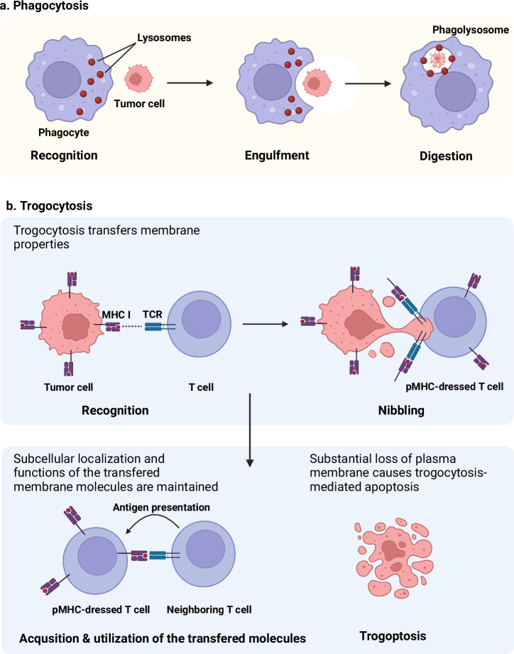
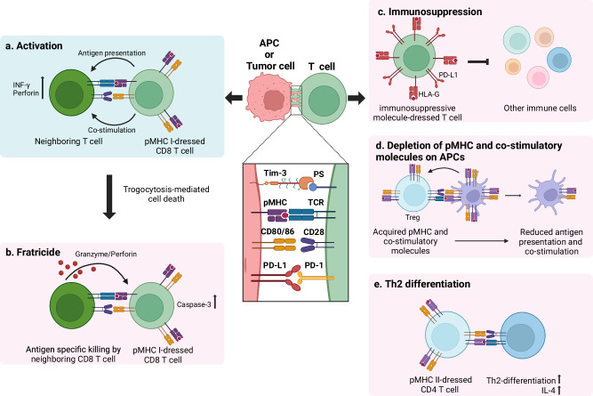
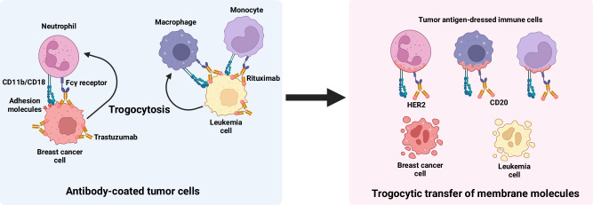
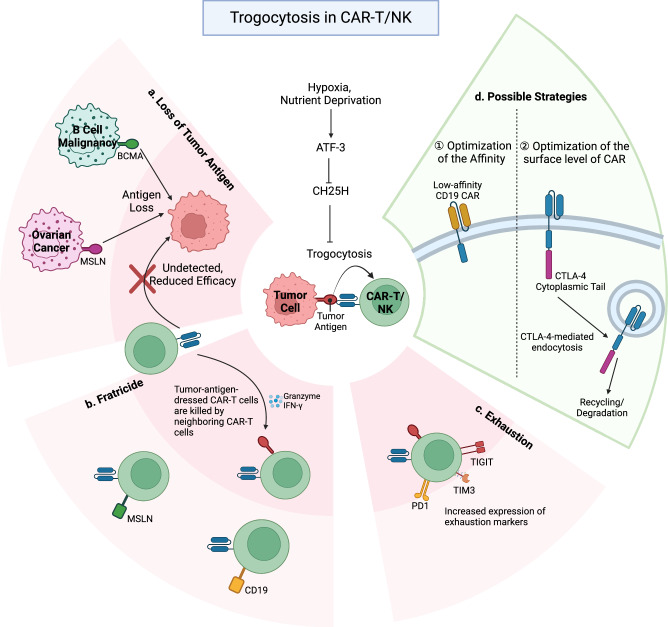
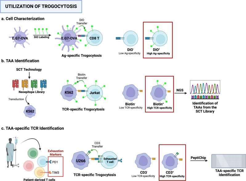
Trogocytosis is a dynamic cellular process characterized by the exchange of the plasma membrane and associated cytosol during cell-to-cell interactions. Unlike phagocytosis, this transfer maintains the surface localization of transferred membrane molecules. For example, CD4 T cells engaging with antigen-presenting cells undergo trogocytosis, which facilitates the transfer of antigen-loaded major histocompatibility complex (MHC) class II molecules from antigen-presenting cells to CD4 T cells. This transfer results in the formation of antigen-loaded MHC class II molecule-dressed CD4 T cells. These "dressed" CD4 T cells subsequently participate in antigen presentation to other CD4 T cells. Additionally, trogocytosis enables the acquisition of immune-regulatory molecules, such as CTLA-4 and Tim3, in recipient cells, thereby modulating their anti-tumor immunity. Concurrently, donor cells undergo plasma membrane loss, and substantial loss can trigger trogocytosis-mediated cell death, termed trogoptosis. This review aims to explore the trogocytosis-mediated transfer of immune regulatory molecules and their implications within the tumor microenvironment to elucidate the underlying mechanisms of immune evasion in cancers.


Conflict of interest statement

Competing interests: The authors declare no competing interests.

Figures

Fig. 1
Fig. 1. Trogocytosis and trogoptosis.
a Phagocytes engulf their target cells, leading to the lysis of the engulfed cells via phagocytosis. b In contrast, trogocytosis is a distinct cellular process in which one cell “nibbles” at another during cell-to-cell contact. This nibbling affects the membrane properties. Importantly, the transferred molecules retain their membrane localization and functions. The nibbling cell acquires functional membrane molecules, whereas the nibbled cell loses portions of its membrane and attaches to the cytosol. Substantial loss of the plasma membrane can lead to apoptosis, a process referred to as trogoptosis.
Fig. 2
Fig. 2. Underlying mechanisms of trogocytosis in T cells.
Trogocytosis is initiated by ligand‒receptor binding, which involves adhesion molecules, such as ICAM-1 and LFA-1, and other cell-specific molecules. T-cell trogocytosis occurs in a TCR affinity-dependent manner. CD28 binding to CD80/86 also triggers trogocytosis. TCR and CD28 stimulation activates the PI3K pathway, leading to cytoskeleton remodeling via actin polymerization, a key mechanism in trogocytosis. PI3K inhibitors, such as wortmannin and LY294002, reduce trogocytosis. Phosphatidylserine on activated T cells binds to Tim-3 on dendritic cells (DCs) to aid the transfer of peptide-loaded MHC molecules to T cells. Regulatory T cells capture CD80/86 on APCs through CTLA-4-mediated trogocytosis by depleting co-stimulatory molecules. CTLA-4 also removes endogenous and trogocytosed CD80/86 on T cells via cis-endocytosis.
Fig. 3
Fig. 3. Consequences of T cell trogocytosis.
The acquisition of immune regulatory molecules by T-cell trogocytosis modulates anti-tumor immunity. a Activation: T cells acquire peptide‒MHC (pMHC) complexes from antigen-presenting cells (APCs) or tumor cells, enabling them to function as APCs. This process leads to the activation of neighboring T cells. b Fratricide: CD8 T cells that acquire pMHC I complexes can become targets of antigen-specific killing by neighboring CD8 T cells, leading to fratricide. c Immunosuppression: The immune regulatory molecules transferred after trogocytosis, such as HLA-G and PD-L1, suppress the reactivity of other immune cells. d Depletion of pMHC and co-stimulatory molecules on APCs: Regulatory T cells (Tregs) can capture co-stimulatory molecules from APCs via CTLA-4-mediated trogocytosis, leading to the depletion of these molecules on APCs. e Th2 differentiation: Trogocytosis of CD4 T cells dressed with pMHC complexes can induce the differentiation of neighboring T cells into Th2 cells, altering the immune response toward a Th2 phenotype.
Fig. 4
Fig. 4. Trogocytosis by NK cells.
Natural killer (NK) cells undergo trogocytosis via interactions between NK cell receptors, such as NKG2D and KIR, and their corresponding ligands. The recruitment of these receptors to the membrane is regulated by the Src kinase pathway; thus, inhibiting Src with PP2 reduces trogocytosis. After trogocytosis, NK cells acquire ligands, such as MICA, Rae-1, or peptide-loaded MHC class I molecules, from tumor cells or antigen-presenting cells (APCs). a MICA- or Rae-1-dressed NK cells are targeted by neighboring NK cells, leading to fratricide. b NK cells acquire immune regulatory molecules such as HLA-G from tumor cells, inhibiting the proliferation and cytotoxicity of other NK cells. c The acquisition of CD9 or PD-1 directly downregulates NK cell reactivity. d NK cells acquire pMHC II and CD80/86 from dendritic cells, presenting antigens to CD4 T cells, but this antigen presentation is less effective than that of professional APCs, leading to reduced T cell responses.
Fig. 5
Fig. 5. Trogocytosis of antibody-coated tumor cells by macrophages, monocytes, and neutrophils.
Neutrophils, macrophages, and monocytes undergo trogocytosis when in contact with antibody-coated tumor cells. Interactions between CD11b/CD18 integrins and adhesion molecules on the tumor cell surface establish a cytotoxic synapse. At this synapse, the Fc gamma receptor (FcγR) binds to trastuzumab bound to HER2, initiating trogocytosis and transferring membrane molecules from breast cancer cells to innate immune cells. A similar process occurs with rituximab-coated leukemia cells. Notably, significant tumor cell membrane loss leads to trogocytosis-mediated apoptosis, known as trogoptosis.
Fig. 6
Fig. 6. Adverse effects of trogocytosis in CAR-T cell therapy.
CAR-T cell trogocytosis disrupts CAR-T cell therapy in the following ways. a CAR-T cells acquire CAR-targeted antigens, such as CD19, BCMA, and MSLN, from leukemia, myeloma, and ovarian cancer cells, respectively, to generate target-free tumor cells. b Tumor antigen-dressed CAR-T cells are attacked by other CAR-T cells, leading to fratricidal cell death. c Prolonged exposure to tumor antigens causes CAR-T cell exhaustion. Trogocytic CAR-T cells, due to their strong antigen affinity, exhibit increased expression of exhaustion markers, such as Tim-3, TIGIT, and PD1. d To counter these adverse effects, low-affinity CD19 CAR-T cells were developed to minimize trogocytosis in the context of B-cell lymphoma. Additionally, fusing the cytoplasmic tail of CTLA-4 to the CAR reduces surface CAR levels, optimizing CAR-T cell therapeutic efficacy.
Fig. 7
Fig. 7. Utilization of trogocytosis.
a Mouse thymoma EL4 cells were labeled with membrane markers, such as 3,3′-dioctadecyl-oxacarbocyanine perchlorate (DiO), and co-cultured with CD8 T cells. As a result, DiO was transferred to CD8 T cells in an antigen-specific manner through trogocytosis. The isolation of DiO+ cells enabled the characterization of antigen-specific CD8 T cells. b Using single-chain trimer technology, a library of tumor-associated antigens (TAAs) was generated and transduced into K562 leukemia cells. Biotin-labeled Jurkat cells were then co-cultured with K562 cells expressing TAAs. Trogocytosis involves the transfer of biotin to target K562 cells. Isolation of biotin+ cells enriched with K562 cells expressing TAAs with high TCR specificity. The selected TAAs were identified via next-generation sequencing (NGS). c Patient-derived T cells expressing exhaustion markers, such as PD-1 or TIM-3, showed enhanced cytotoxicity against target cells, indicating high antigen specificity. After these PD-1+ TIM-3+ T cells were co-cultured with U266 cells, trogocytosis transferred CD3 from the T cells to the U266 cells. Isolation of CD3+ U266 cells after co-culture, followed by TCR identification, can reveal TAA-specific TCRs via the PeptiChip technique.

References

    1. Cone, R. E., Sprent, J. & Marchalonis, J. J. Antigen-binding specificity of isolated cell-surface immunoglobulin from thymus cells activated to histocompatibility antigens. Proc. Natl Acad. Sci. USA69, 2556–2560 (1972). - PMC - PubMed
    1. Hudson, L., Sprent, J., Miller, J. F. A. P. & Playfair, J. H. L. B cell-derived immunoglobulin on activated mouse T lymphocytes. Nature251, 60–62 (1974). - PubMed
    1. Huang, J.-F. et al. TCR-mediated internalization of peptide-MHC complexes acquired by T cells. Science286, 952–954 (1999). - PubMed
    1. Joly, E. & Hudrisier, D. What is trogocytosis and what is its purpose? Nat. Immunol.4, 815–815 (2003). - PubMed
    1. Shin, J. H. et al. Colon cancer cells acquire immune regulatory molecules from tumor-infiltrating lymphocytes by trogocytosis. Proc. Natl Acad. Sci. USA118, e2110241118 (2021). - PMC - PubMed