

A case of subcutaneous metastatic malignant melanoma of the left medial ankle: a case report and review of literature
- PMID: 39741330
- PMCID: PMC11689692
- DOI: 10.1186/s13256-024-04908-2
A case of subcutaneous metastatic malignant melanoma of the left medial ankle: a case report and review of literature
Abstract
Background: Although rare, melanoma confined to the dermis or subcutaneous tissue without evidence of a primary cutaneous site should provoke consideration of melanoma of unknown primary. This diagnosis carries a favorable prognosis when compared with cutaneous metastatic melanoma. Several hypotheses have been proposed for how melanoma of unknown primary develops, two of which were considered in our patient case: (1) spontaneous regression of the primary tumor following metastasis or (2) the traumatic implantation of ectopic melanocytic cells in other tissues, such as the subcutaneous tissue. Although not a true example of melanoma of unknown primary, our case is still noteworthy as it represents a unique instance of melanoma presenting subcutaneously from trauma to a preexisting epidermal nevus.
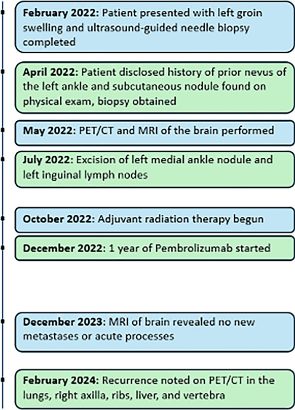
Case presentation: We present the case of a 66-year-old non-Hispanic Caucasian male who initially sought evaluation for a nontender lump of the left groin. Ultrasound-guided needle biopsy demonstrated stage III malignant melanoma. Upon further history taking, it was discovered that he had a nevus of the left medial ankle that was subjected to traumatic removal. He later developed a subcutaneous nodule at the same site. Positron emission tomography scan results supported the histopathologic findings which demonstrated invasive melanoma centered in the subcutaneous tissue without an epidermal component. Following left inguinal lymph node dissection, the patient received adjuvant immunotherapy and radiation to the left inguinal area. At 6 months following completion of therapy, metastases were identified in the lungs, vertebra, ribs, and liver. The patient is currently receiving immunotherapy with ipilimumab-nivolumab.
Conclusion: As our patient did not have a readily apparent primary epidermal melanoma site at presentation, consideration was given as to whether this case may represent a melanoma of unknown primary, as originally defined by Das Gupta. This case does not meet the proposed criteria, however, as the patient reported a preexisting nevus in the area that was subjected to traumatic removal. Instead, we postulate that this trauma allowed for implantation of melanocytes into the subcutaneous tissue that later resulted in a malignant melanoma.
Keywords: Case report; Malignant melanoma; Melanoma of unknown primary; Metastatic melanoma; Subcutaneous melanoma; Surgical excision; Trauma.
© 2024. This is a U.S. Government work and not under copyright protection in the US; foreign copyright protection may apply.
Conflict of interest statement
Declarations. Ethics approval and consent to participate: A Case Report Summary and Acknowledgement application was submitted to ProMedica institutional review board (PHS IRB) for review. The PHS IRB determined that the case report was not considered human subject research and that further IRB review would not be required. No patient identifiers are included in this manuscript. Consent for publication: Written informed consent was obtained from the patient for publication of this case report and any accompanying images. A copy of the written consent is available for review by the Editor-in-Chief of this journal. Competing interests: The authors declare that they have no competing interests.
Figures

Similar articles
-
Metastatic melanoma from an unknown primary site presenting as skin-colored nodules and multiple visceral involvement.Skinmed. 2012 Nov-Dec;10(6):396-9. Skinmed. 2012. PMID: 23346671
-
Unmasking of intracranial metastatic melanoma during ipilimumab/nivolumab therapy: case report and literature review.BMC Cancer. 2018 May 9;18(1):549. doi: 10.1186/s12885-018-4470-y. BMC Cancer. 2018. PMID: 29743050 Free PMC article. Review.
-
Role of In Vivo Reflectance Confocal Microscopy in the Analysis of Melanocytic Lesions.Acta Dermatovenerol Croat. 2018 Apr;26(1):64-67. Acta Dermatovenerol Croat. 2018. PMID: 29782304 Review.
-
Giant metastatic malignant melanoma with an unknown primary site.J Dermatol. 1994 Jun;21(6):442-6. doi: 10.1111/j.1346-8138.1994.tb01771.x. J Dermatol. 1994. PMID: 8064010
-
Primary Dermal Melanoma: A Case Report.Am J Dermatopathol. 2022 Mar 1;44(3):212-214. doi: 10.1097/DAD.0000000000002099. Am J Dermatopathol. 2022. PMID: 34726186
References
-
- Scott JF, Gerstenblith MR. Melanoma of unknown primary. Noncutaneous Melanoma: Brisbane (AU); 2018.
-
- González-de Arriba M, Bordel-Gómez MT, Solera JC, Sánchez-Estella J. Primary dermal melanoma: a case report and a review of the literature. Actas Dermosifiliogr. 2013;104(6):518–22. 10.1016/j.ad.2011.12.020. - PubMed
-
- Keung EZ, Balch CM, Thompson JF, Kirkwood JM, Scolyer RA, Sondak VK, et al. Melanoma prognosis and staging. In: Balch CM, Atkins MB, Garbe C, Gershenwald JE, Halpern AC, Kirkwood JM, et al., editors. Cutaneous Melanoma. Cham: Springer International Publishing; 2020. p. 271–97.
-
- Song Y, Karakousis GC. Melanoma of unknown primary. J Surg Oncol. 2019;119(2):232–41. 10.1002/jso.25302. - PubMed
Publication types
MeSH terms
LinkOut - more resources
Full Text Sources
Medical
Research Materials
