. 2025 Feb 5;33(2):786-804.
doi: 10.1016/j.ymthe.2024.12.039. Epub 2024 Dec 30.

Modulation of pain sensitivity by Ascl1- and Lhx6-dependent GABAergic neuronal function in streptozotocin diabetic mice

Affiliations

Modulation of pain sensitivity by Ascl1- and Lhx6-dependent GABAergic neuronal function in streptozotocin diabetic mice

Sung-Min Hwang et al. Mol Ther. .

Abstract

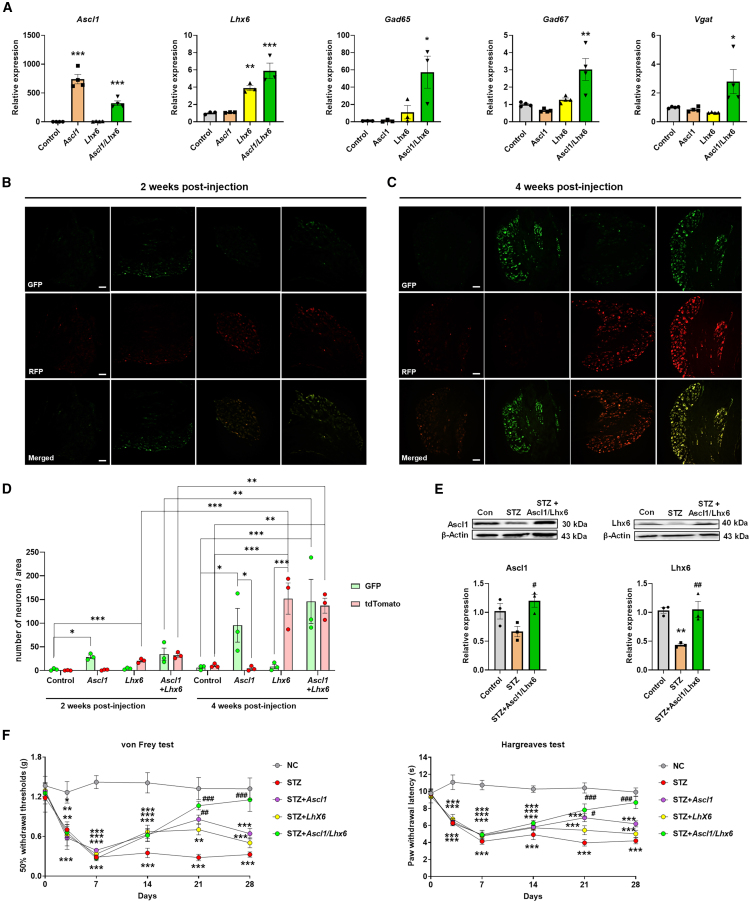
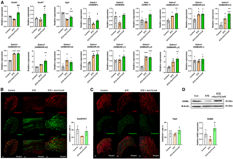
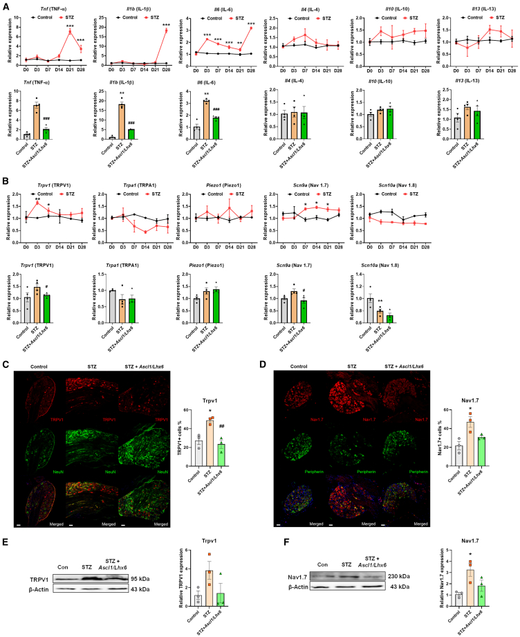
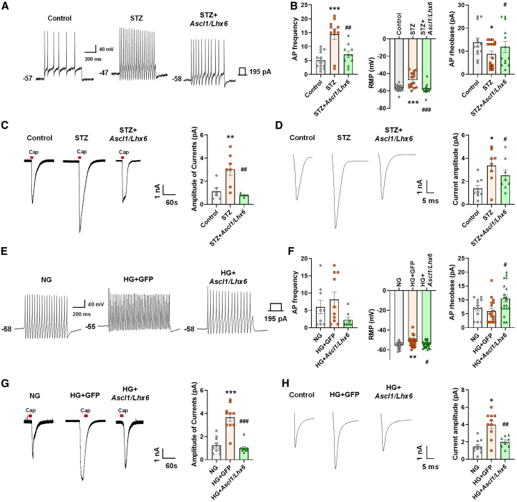
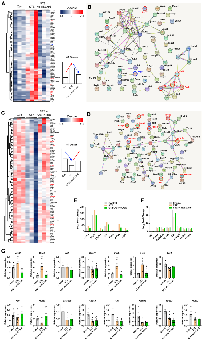
Painful diabetic neuropathy commonly affects the peripheral nervous system in individuals with diabetes. However, the pathological processes and mechanisms underlying diabetic neuropathic pain remain unclear. We aimed to identify the overall profiles and screen for genes potentially involved in pain mechanisms using transcriptome analysis of the dorsal root ganglion of diabetic mice treated with streptozotocin (STZ). Using RNA sequencing, we identified differentially expressed genes between streptozotocin-treated diabetic mice and controls, focusing on altered GABAergic neuron-related genes and inflammatory pathways. Behavioral and molecular analyses revealed a marked reduction in GABAergic neuronal markers (GAD65, GAD67, VGAT) and increased pro-inflammatory cytokines (TNF-α, IL-1β, IL-6) in the diabetic group compared with controls. Intrathecal administration of lentiviral vectors expressing transcription factors Ascl1 and Lhx6 reversed pain hypersensitivity and restored normal expression of GABAergic genes and inflammatory mediators. Protein-protein interaction network analysis revealed five key proteins influenced by Ascl1 and Lhx6 treatment, including those in the JunD/FosB/C-fos signaling pathway. These findings suggest that Ascl1 and Lhx6 mitigate diabetic neuropathic pain by modulating GABAergic neuronal function, pro-inflammatory responses, and pain-related channels (TRPV1, Nav1.7). These results provide a basis for developing transcription factor-based therapies targeting GABAergic neurons for diabetic neuropathic pain relief.

Keywords: GABAergic neuron; RNA sequencing; diabetic neuropathic pain; dorsal root ganglion; transcription factor.


Conflict of interest statement

Declaration of interests The authors declare no competing interests.

Figures

None
Graphical abstract
Figure 1
Figure 1
RNA sequencing transcriptional profiling and identification of abnormal pain-related gene expression in the DRG of diabetic mice (A) Timelines of the in vivo experimental design. (B) Mechanical and thermal tests in STZ-induced diabetic mice. (C) Hierarchical clustering analyses of DEGs. (D) Volcano plot of DEGs. (E) The number of upregulated and downregulated DEGs. (F) GO analysis of DEGs in the control versus STZ group. Data are reported as mean ± standard errors of the mean (n = 5). ∗p < 0.05, ∗∗∗p < 0.001 via the Bonferroni multiple comparisons test by two-way analysis of variance versus control. Data are shown for D0, D3, D7, D14, D21, and D28. STZ, streptozotocin.
Figure 2
Figure 2
Downregulation of GABAergic neuron-specific genes and their transcription factors in the DRG with diabetes (A) Relative mRNA expression level of GABAergic neuron-specific genes. (B) Relative mRNA expression level of transcription factors in controlling the GABAergic neuron subtype identity. Data are reported as mean ± standard errors of the mean (n = 5). ∗p < 0.05, ∗∗p < 0.01, ∗∗∗p < 0.001, Dunnett’s multiple comparisons tests by one-way analysis of variance versus day 0. Data are shown for D0, D3, D7, D14, D21, and D28. STZ, streptozotocin.
Figure 3
Figure 3
Synergistic effects of Ascl1 and Lhx6 through transgene expression alleviate mechanical allodynia and thermal hyperalgesia in diabetic neuropathy (A) Relative mRNA expression levels of GABAergic neuron-specific genes after treatment with Ascl1 and Lhx6 (n = 4). (B and C) Representative images showing GFP (green) and dTomato (red) fluorescence in mouse DRG tissues 2 weeks post-injection (B) and 4 weeks post-injection (C) of lentiviral vectors: empty vector (control), Lenti-hSyn-mAscl-P2A-EGFP (Ascl1), Lenti-hSyn-mLhx6-P2A-dTomato (Lhx6), and co-injection of Lenti-hSyn-mAscl-P2A-EGFP and Lenti-hSyn-mLhx6-P2A-dTomato (Ascl1+Lhx6). Scale bars, 50 μm. (D) Quantitative analysis of the number of EGFP- and tdTomato-positive cells per unit area in DRG tissues (n = 3). These data demonstrate the successful expression of Ascl1 and Lhx6 in DRG tissues and highlight the increased expression efficiency with time. (E) Relative protein expression levels of Ascl1 and Lhx6 in the three groups; β-actin was used as an internal control (n = 3). (F) Mechanical and thermal tests, using the von Frey test and Hargreaves test, respectively, in STZ-induced diabetic mice demonstrating the inhibitory effects of Ascl1 and Lhx6 on pain sensitivity (n = 5). Data are reported as mean ± standard error of the mean. ∗p < 0.05, ∗∗p < 0.01, ∗∗∗p < 0.001, Tukey’s multiple comparisons tests by two-way analysis of variance versus the naive group; #p < 0.05, ##p < 0.01, ###p < 0.001, versus the STZ diabetic control group. The data of non-significant points are not marked. STZ, streptozotocin.
Figure 4
Figure 4
Regulatory effects of Ascl1 and Lhx6 on GABAergic neuron-specific genes in the DRG of diabetic mice (A) Relative mRNA expression level of GABA-related genes in the three groups. (B and C) Relative protein expression level of GABAergic neuron-specific protein, Gad65/67, and Vgat in three groups. (D) Relative protein expression level of GABA in the three groups; β-actin was used as an internal control. Data are reported as mean ± standard error of the mean (n = 3). ∗p < 0.05, ∗∗∗p < 0.001, Tukey’s multiple comparisons tests by two-way analysis of variance versus the naive group; #p < 0.05, ##p < 0.01, ###p < 0.001, versus the STZ diabetic control group. The data of non-significant points are not marked. STZ, streptozotocin.
Figure 5
Figure 5
Regulatory function of Ascl1 and Lhx6 on GABAergic neuron-specific genes, pro-inflammatory and anti-inflammatory cytokines and pain-related channels in the DRG of diabetic mice (A) Relative mRNA expression level of pro- and anti-inflammatory cytokine-related factors in the three groups. (B) Relative mRNA expression level of pain-related channels in the three groups. (C and D) Relative protein expression levels of TRPV1 and Nav1.7 in the three groups. (E and F) Relative protein expression level of GABA in the three groups; β-actin was used as an internal control. Data are reported as mean ± standard error of the mean (n = 3). ∗p < 0.05, ∗∗∗p < 0.001, Tukey’s multiple comparisons tests by two-way analysis of variance versus the naive group; #p < 0.05, ###p < 0.001, versus the STZ diabetic control group. STZ, streptozotocin.
Figure 6
Figure 6
Regulatory effects of Ascl1 and Lhx6 on neuronal hyperexcitability, TRPV1, and Navs in the DRG of diabetic mice (A) Representative example of the AP frequency, RMP, and minimum current (rheobase) in the three groups in vivo. (B) Comparison of the AP frequency, RMP, and minimum current (rheobase) in the three groups in vivo. (C) Representative example (left) and comparison (right) of TRPV1 activation in the DRG neurons in the three groups in vivo. (D) Representative example (left) and comparison (right) of the transient Na+ current in small-sized DRG neurons in the three groups in vivo. (E) Representative example of the AP frequency, RMP, and minimum current (rheobase) in the three groups in vitro. (F) Comparison of the AP frequency, RMP, and minimum current (rheobase) in the three groups in vitro. (G) Representative example (left) and comparison (right) of TRPV1 activation in the DRG neurons in the three groups in vitro. (H) Representative example (left) and comparison (right) of transient Na+ current activation in small-sized DRG neurons in the three groups in vitro. Data are reported as mean ± standard error of the mean. ∗p < 0.05, ∗∗p < 0.01, ∗∗∗p < 0.001, Tukey’s multiple comparisons tests by two-way analysis of variance versus the naive group; #p < 0.05, ##p < 0.01, ###p < 0.001, versus the STZ diabetic control group. STZ, streptozotocin.
Figure 7
Figure 7
Identification of candidate genes influenced by Ascl1 and Lhx6 in the DRG of diabetic mice (A–D) Hierarchical clustering heatmap of DEGs and the network generated by STRING database analysis in the three groups. (E and F) Of the genes identified in (A), 15 are significantly expressed. (G) Relative mRNA expression levels of 15 candidate genes in the 3 groups. Data are reported as mean ± standard error of the mean (n = 3). ∗p < 0.05, ∗∗p < 0.01, Tukey’s multiple comparisons tests by two-way analysis of variance versus the naive group; #p < 0.05, ##p < 0.01, versus the STZ diabetic control group. STZ, streptozotocin.
Figure 8
Figure 8
Identification of five proteins in the DRG of diabetic mice (A) Expression changes in FoxK1, C-Fos, FosB, JunD, and GNG3 via immunoblot analysis in the three groups; β-actin was used as an internal control (n = 3). (B and C) Network generated by STRING database analysis using the DEGs from (A). (D) Proposed schematic of the molecular mechanisms underlying pain regulation in the three conditions (no pain, persistent pain sensation, and reduced pain sensation) through the overexpression of Ascl1/Lhx6. Data are reported as mean ± standard error of the mean. ∗p < 0.05, ∗∗p < 0.01, Tukey’s multiple comparisons tests by two-way analysis of variance versus the naive group; #p < 0.05, ##p < 0.01, versus the STZ diabetic control group. STZ, streptozotocin.

References

    1. Pop-Busui R., Ang L., Boulton A.J.M., Feldman E.L., Marcus R.L., Mizokami-Stout K., Singleton J.R., Ziegler D. American Diabetes Association; 2022. Diagnosis and Treatment of Painful Diabetic Peripheral Neuropathy; pp. 1–32. Arlington, VA. - DOI - PubMed
    1. Schreiber A.K., Nones C.F., Reis R.C., Chichorro J.G., Cunha J.M. Diabetic neuropathic pain: Physiopathology and treatment. World J. Diabetes. 2015;6:432–444. doi: 10.4239/wjd.v6.i3.432. - DOI - PMC - PubMed
    1. Chung J.M., Chung K. Importance of hyperexcitability of DRG neurons in neuropathic pain. Pain Pract. 2002;2:87–97. doi: 10.1046/j.1533-2500.2002.02011.x. - DOI - PubMed
    1. Reyes-Pardo H., Sanchez-Herrera D.P., Santillan M. On the effects of diabetes mellitus on the mechanical properties of DRG sensory neurons and their possible relation with diabetic neuropathy. Phys. Biol. 2022;19:1–4. doi: 10.1088/1478-3975/ac6722. - DOI - PubMed
    1. Millan M.J. Descending control of pain. Prog. Neurobiol. 2002;66:355–474. doi: 10.1016/s0301-0082(02)00009-6. - DOI - PubMed

MeSH terms

Substances

LinkOut - more resources