. 2024 Dec 17:5:1466281.
doi: 10.3389/fragi.2024.1466281. eCollection 2024.

The aldose reductase inhibitors AT-001, AT-003 and AT-007 attenuate human keratinocyte senescence

Affiliations

The aldose reductase inhibitors AT-001, AT-003 and AT-007 attenuate human keratinocyte senescence

Gautham Yepuri et al. Front Aging. .

Abstract

Human skin plays an important role protecting the body from both extrinsic and intrinsic factors. Skin aging at cellular level, which is a consequence of accumulation of irreparable senescent keratinocytes is associated with chronological aging. However, cell senescence may occur independent of chronological aging and it may be accelerated by various pathological conditions. Recent studies have shown that oxidative stress driven keratinocyte senescence is linked to the rate limiting polyol pathway enzyme aldose reductase (AR). Here we investigated the role of three novel synthetic AR inhibitors (ARIs) AT-001, AT-003 and AT-007 in attenuating induced skin cell senescence, in primary normal human keratinocytes (NHK cells), using three different senescence inducing agents: high glucose (HG), hydrogen peroxide (H2O2) and mitomycin-c (MMC). To understand the efficacy of ARIs in reducing senescence, we have assessed markers of senescence, including SA-β-galactosidase activity, γ-H2AX foci, gene expression of CDKN1A, TP53 and SERPINE1, reactive oxygen species generation and senescence associated secretory phenotypes (SASP). Strikingly, all three ARIs significantly inhibited the assessed senescent markers, after senescence induction. Our data confirms the potential role of ARIs in reducing NHK cell senescence and paves the way for preclinical and clinical testing of these ARIs in attenuating cell aging and aging associated diseases.

Keywords: aldose reductase; aldose reductase inhibitors; oxidative stress; senescence; skin cell aging.


Conflict of interest statement

Authors RP, SS, and AW were employed by Applied Therapeutics Inc. The remaining authors declare that the research was conducted in the absence of any commercial or financial relationships that could be construed as a potential conflict of interest. The authors declare that this study received funding from Applied Therapeutics Inc. The funder had the following involvement in the study: editing the manuscript.

Figures

FIGURE 1
FIGURE 1
Senescence induction upregulates AR activity. AR activity measured by colorimetric assay in human keratinocytes either treated with (A) 10 uM H2O2 or Vehicle (Veh- dH2O) (B) 25 mM high glucose (HG) or normal glucose (5 mM) (NG) for 48 h (C) 200 nM Mitomycin-C (MMC) for 24 h compared to Veh (DMSO) and (D) 0.2 nM AT-001 for 48 h vs. Veh (1:5,000 dilution DMSO in PBS). Data are presented as the mean ± SEM. n = 4 per group obtained from two consecutive experiments. Unpaired t-test was implemented for comparison between two groups. p* < 0.05 vs. Veh or NG.
FIGURE 2
FIGURE 2
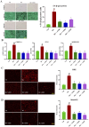
ARIs prevent HG induced senescence phenotype. Human keratinocytes were treated with 25 mM HG or normal condition medium NG. (A) SA-β-gal staining was performed to assess positive senescent cells. (B) qPCR analysis for confirming gene expression of senescent markers CDKN1A, TP53 and SERPINE1. (C) Skin Cells stained with 5 µM DiHydroethidium (DHE) dye for 30 min to detect cytosolic superoxide and (D) cells stained with 5 µM MitoSOX dye for 10 min to detect mito superoxide. Data are presented as the mean ± SEM. n = 4 per group obtained from two consecutive experiments. Ordinary one-way ANOVA with Tukey’s multiple comparisons test was implemented for comparison between groups. p* < 0.05 vs. NG. p# < 0.05 vs. Veh.
FIGURE 3
FIGURE 3
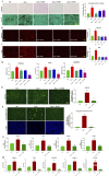
ARIs prevent H2O2 induced senescence phenotype. Human keratinocytes were treated with 10 µM H2O2 or Veh (dH2O) and ARIs for 48 h. (A) SA-β-gal staining was performed to assess positive senescent cells. (B) Skin Cells stained with 5 µM DiHydroethidium (DHE) dye for 30 min to detect cytosolic superoxide and (C) cells stained with 5 µM MitoSOX dye for 10 min to detect mito superoxide. (D) qPCR analysis for confirming gene expression of senescent markers CDKN1A, TP53 and SERPINE1. (E) Gamma H2A.X Staining and (F) CellEvent™ Senescence Green Detection staining in human keratinocytes treated with Veh (dH2O), 400 µM H2O2 for 1 h followed by assessment of cells at day 10 with or without ARI AT-001. (G) ELISA to measure cytokine release in condition medium collected at day 10 and (H) qPCR analysis for confirming gene expression of senescent markers CDKN1A, TP53 and SERPINE1 CDKN2A and LMNB1. Data are presented as the mean ± SEM. n = 4 per group obtained from two consecutive experiments. Ordinary one-way ANOVA with Tukey’s multiple comparisons test was implemented for comparison between groups. p* < 0.05 vs. H2O2. p# < 0.05 vs. Veh.
FIGURE 4
FIGURE 4
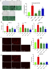
ARIs prevent MMC induced senescence phenotype. Human keratinocytes were treated with 200 nM MMC or Veh (DMSO) and ARIs for 24 h. (A) SA-β-gal staining was performed to assess positive senescent cells. (B) qPCR analysis for confirming gene expression of senescent markers CDKN1A, TP53 and SERPINE1. (C) Skin Cells stained with 5 µM DiHydroethidium (DHE) dye for 30 min to detect cytosolic superoxide and (D) cells stained with 5 µM MitoSOX dye for 10 min to detect mito superoxide. Data are presented as the mean ± SEM. n = 4 per group obtained from two consecutive experiments. Ordinary one-way ANOVA with Tukey’s multiple comparisons test was implemented for comparison between groups. p* < 0.05 vs. MMC. p# < 0.05 vs. Veh.
SCHEME 1
SCHEME 1
Schematic representation showing AR activity increases senescent markers thereby promoting skin cell aging which can be inhibited by treatment with ARIs.

References

    1. Ananthakrishnan R., Kaneko M., Hwang Y. C., Quadri N., Gomez T., Li Q., et al. (2009). Aldose reductase mediates myocardial ischemia-reperfusion injury in part by opening mitochondrial permeability transition pore. Am. J. Physiol. Heart Circ. Physiol. 296 (2), H333–H341. 10.1152/ajpheart.01012.2008 - DOI - PMC - PubMed
    1. Argyropoulos A. J., Robichaud P., Balimunkwe R. M., Fisher G. J., Hammerberg C., Yan Y., et al. (2016). Alterations of dermal connective tissue collagen in diabetes: molecular basis of aged-appearing skin. PLoS One 11 (4), e0153806. 10.1371/journal.pone.0153806 - DOI - PMC - PubMed
    1. Chen C., Zhou M., Ge Y., Wang X. (2020). SIRT1 and aging related signaling pathways. Mech. Ageing Dev. 187, 111215. 10.1016/j.mad.2020.111215 - DOI - PubMed
    1. Cosgrove M. C., Franco O. H., Granger S. P., Murray P. G., Mayes A. E. (2007). Dietary nutrient intakes and skin-aging appearance among middle-aged American women. Am. J. Clin. Nutr. 86 (4), 1225–1231. 10.1093/ajcn/86.4.1225 - DOI - PubMed
    1. Danby F. W. (2010). Nutrition and aging skin: sugar and glycation. Clin. Dermatol. 28 (4), 409–411. 10.1016/j.clindermatol.2010.03.018 - DOI - PubMed

LinkOut - more resources