. 2024 Dec 4;57(24):11738-11752.
doi: 10.1021/acs.macromol.4c02016. eCollection 2024 Dec 24.

RAFT Dispersion Polymerization of 2-Hydroxyethyl Methacrylate in Non-polar Media

Affiliations

RAFT Dispersion Polymerization of 2-Hydroxyethyl Methacrylate in Non-polar Media

Priyanka Chohan et al. Macromolecules. .

Abstract

We report the reversible addition-fragmentation chain transfer (RAFT) dispersion polymerization of 2-hydroxyethyl methacrylate (HEMA) in n-dodecane using a poly(lauryl methacrylate) (PLMA) precursor at 90 °C. This formulation is an example of polymerization-induced self-assembly (PISA), which leads to the formation of a colloidal dispersion of spherical PLMA-PHEMA nanoparticles at 10-20% w/w solids. PISA syntheses involving polar monomers in non-polar media have been previously reported but this particular system offers some unexpected and interesting challenges in terms of both synthesis and characterization. First, GPC analysis requires chemical derivatization of the pendent hydroxyl groups in the PHEMA block using excess acetyl chloride to ensure that both blocks are fully soluble in chloroform. Second, DLS, TEM and 1H NMR spectroscopy studies of the periodically sampled polymerizing mixture indicate the transient formation of anomalously large, colloidally unstable aggregates at around 50% conversion, which approximately corresponds to the maximum rate of polymerization. Remarkably, such aggregates immediately break up to form well-defined nanoparticles, which remain colloidally stable at the end of the HEMA polymerization. Moreover, depending on the target degree of polymerization (DP) for the PHEMA block, TEM studies typically indicate bimodal particle size distributions for PLMA-PHEMA nanoparticles prepared using a one-shot batch protocol. This is attributed to a side-reaction between HEMA monomer and the dithiobenzoate-based RAFT agent. Fortunately, this problem can be prevented by conducting such PISA syntheses under monomer-starved conditions by continuous addition of the HEMA monomer using a syringe pump. Alternatively, unimodal spheres can also be produced via adding HEMA in multiple batches. This PISA formulation has been optimized to produce monomodal particle size distributions while targeting a PHEMA DP of up to 1000 at the maximum possible copolymer concentration. Finally, time-resolved small-angle X-ray scattering (SAXS) studies indicate the rapid formation of well-defined near-monodisperse spheres when targeting PLMA14-PHEMA50 nanoparticles.


Conflict of interest statement

The authors declare no competing financial interest.

Figures
Scheme 1
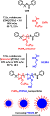
Scheme 1. One-Pot Synthesis of a Poly(lauryl methacrylate) (PLMA) Precursor via RAFT Solution Polymerization in n-Dodecane Using Cumyl Dithiobenzoate (CDB) at 90 °C, Immediately Followed by the RAFT Dispersion Polymerization of 2-Hydroxyethyl Methacrylate (HEMA) in n-Dodecane at 90 °C
Scheme 2
Scheme 2. Esterification of PLMA14–PHEMAy Diblock Copolymer in n-Dodecane at 25 °C Using Excess Acetyl Chloride ([CH3COCl]/[HEMA] Molar Ratio = 1.5) to Enable Chloroform GPC Analysis
Figure 1
Figure 1
(a) Chloroform GPC curves [vs a series of near-monodisperse poly(methyl methacrylate) calibration standards, which incur a systematic error in the Mn data] recorded using a refractive index detector for a PLMA14 precursor (black dashed curve) prepared by RAFT solution polymerization of LMA in n-dodecane at 80% w/w solids, and ten corresponding PLMA14–PHEMAy diblock copolymers prepared by RAFT dispersion polymerization of HEMA in n-dodecane at 90 °C when targeting 20% w/w solids and y = 25 – 150. (b) Linear relationship between GPC Mn (blue circles) and actual PHEMA DP (as determined by 1H NMR studies) observed for the same series of PLMA14–PHEMAy diblock copolymers. The corresponding Mw/Mn (red squares) data are also shown.
Figure 2
Figure 2
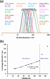
(a) DLS particle size distributions recorded for 0.1% w/w dispersions of a series of PLMA14–PHEMAy nanoparticles (y = 25–149) prepared by RAFT dispersion polymerization of HEMA in n-dodecane at 90 °C when targeting 20% w/w solids. (b) Relationship between the apparent z-average diameter and the PHEMA DP for the same series of nanoparticles.
Figure 3
Figure 3
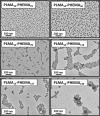
Representative TEM images recorded for selected PLMA14–PHEMAy nanoparticles (where y = 25, 50, 78, 119, 127 and 149).
Figure 4
Figure 4
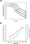
(a) SAXS patterns recorded for 1.0% w/w diblock copolymer dispersions in n-dodecane (PLMA14–PHEMA25, black squares; PLMA14–PHEMA50, green circles; PLMA14–PHEMA78, blue triangles; and PLMA14–PHEMA119, red diamonds) fitted using a well-known spherical micelle model (gray curves). For clarity, these SAXS patterns are offset by an arbitrary multiplication factor. (b) Variation in volume-average core diameter (red data) and mean aggregation number (blue data) with PHEMA DP for the same four examples of PLMA14–PHEMA25–119 nanoparticles.
Figure 5
Figure 5
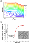
(a) Time-resolved SAXS patterns recorded during the RAFT dispersion polymerization of HEMA at 90 °C when targeting 20% w/w solids in n-dodecane. For clarity, each SAXS pattern has been scaled by an arbitrary factor. (b) Plot of volume-average core diameter vs reaction time for the same polymerization. This kinetic experiment had a dead time of 2.37 min (see Figure S1) and the first primary minimum was observed after 10.68 min. Inset: representative TEM image recorded for the final PLMA14–PHEMA48 nanoparticles.
Figure 6
Figure 6
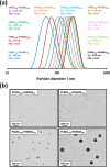
(a) DLS particle size distributions recorded for 0.1% w/w dispersions of PLMA196–PHEMAy (y = 95–990) nanoparticles. (b) Representative TEM images recorded for selected PLMA196–PHEMAy nanoparticles (y = 95, 196, 294 or 891).
Scheme 3
Scheme 3. Postulated Side-Reaction between HEMA and CDB at 90 °C
1H NMR spectroscopy and LC–MS analysis (see Figure S4) suggest that this HEMA/CDB adduct is formed on heating a 10:1 HEMA/CDB mixture for 3 h in the absence of any initiator. A similar side-reaction is proposed to occur between HEMA and the dithiobenzoate-capped copolymer chains at 90 °C within the monomer-swollen PLMA–PHEMA nanoparticles formed during the PISA syntheses reported herein.
Figure 7
Figure 7
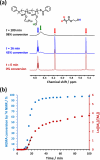
(a) Selected partial 1H NMR spectra recorded during the RAFT dispersion polymerization of HEMA in n-dodecane at 90 °C when targeting PLMA196–PHEMA1000 nanoparticles at 10% w/w solids: t = 0 min (red curve), t = 16 min (blue curve) and t = 100 min (black curve). (b) Conversion vs time curve (blue data) and corresponding semilogarithmic plot (red data) calculated for the same PISA formulation.
Figure 8
Figure 8
Evolution in z-average diameter (blue data) and DLS polydispersity (red data) over time recorded for aliquots periodically extracted from the reaction mixture when targeting PLMA196–PHEMA1000 nanoparticles using the one-shot batch protocol at 90 °C in n-dodecane at 10% w/w solids. Inset: digital photographs recorded to illustrate the physical appearance of aliquots extracted after 14, 16, and 18 min.
Figure 9
Figure 9
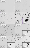
Representative TEM images recorded for aliquots extracted after 8, 10, 14, 16, 18, 20, 25, and 100 min, respectively when targeting PLMA196–PHEMA1000 nanoparticles using the one-shot batch protocol at 90 °C in n-dodecane at 10% w/w solids.
Figure 10
Figure 10
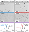
(a) DLS particle size distributions recorded for 0.1% w/w dispersions of PLMA196–PHEMA1000 nanoparticles prepared at 10% w/w solids in n-dodecane with HEMA monomer addition either by the one-shot batch protocol or by sequential addition of two, four or eight equal batches. (b) Representative TEM images recorded for the same nanoparticles.
Figure 11
Figure 11
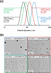
Representative TEM images recorded when targeting PLMA196–PHEMA400 and PLMA196–PHEMA1000 nanoparticles prepared by the one-shot batch protocol (black) and monomer-starved addition of HEMA monomer (blue and red). The corresponding DLS particle size distributions recorded for 0.1% w/w dispersions of PLMA196–PHEMA400 and PLMA196–PHEMA1000 nanoparticles prepared via one-shot batch addition or monomer-starved addition of HEMA are also shown (purple panels).

References

    1. Charleux B.; Delaittre G.; Rieger J.; D’Agosto F. Polymerization-Induced Self-Assembly: From Soluble Macromolecules to Block Copolymer Nano-Objects in One Step. Macromolecules 2012, 45, 6753–6765. 10.1021/ma300713f. - DOI
    1. Penfold N. J. W.; Yeow J.; Boyer C.; Armes S. P. Emerging Trends in Polymerization-Induced Self-Assembly. ACS Macro Lett. 2019, 8, 1029–1054. 10.1021/acsmacrolett.9b00464. - DOI - PubMed
    1. D’Agosto F.; Rieger J.; Lansalot M. RAFT-Mediated Polymerization-Induced Self-Assembly. Angew. Chem., Int. Ed. 2020, 59, 8368–8392. 10.1002/anie.201911758. - DOI - PubMed
    1. Wang X.; An Z. New Insights into RAFT Dispersion Polymerization-Induced Self-Assembly: From Monomer Library, Morphological Control, and Stability to Driving Forces. Macromol. Rapid Commun. 2019, 40, 1800325.10.1002/marc.201800325. - DOI - PubMed
    1. Liu C.; Hong C. Y.; Pan C. Y. Polymerization Techniques in Polymerization-Induced Self-Assembly (PISA). Polym. Chem. 2020, 11, 3673–3689. 10.1039/D0PY00455C. - DOI

LinkOut - more resources