. 2024 Dec 7:2024:5538740.
doi: 10.1155/cdr/5538740. eCollection 2024.

Dehydroevodiamine Alleviates Doxorubicin-Induced Cardiomyocyte Injury by Regulating Neuregulin-1/ErbB Signaling

Affiliations

Dehydroevodiamine Alleviates Doxorubicin-Induced Cardiomyocyte Injury by Regulating Neuregulin-1/ErbB Signaling

Song Jie et al. Cardiovasc Ther. .

Abstract

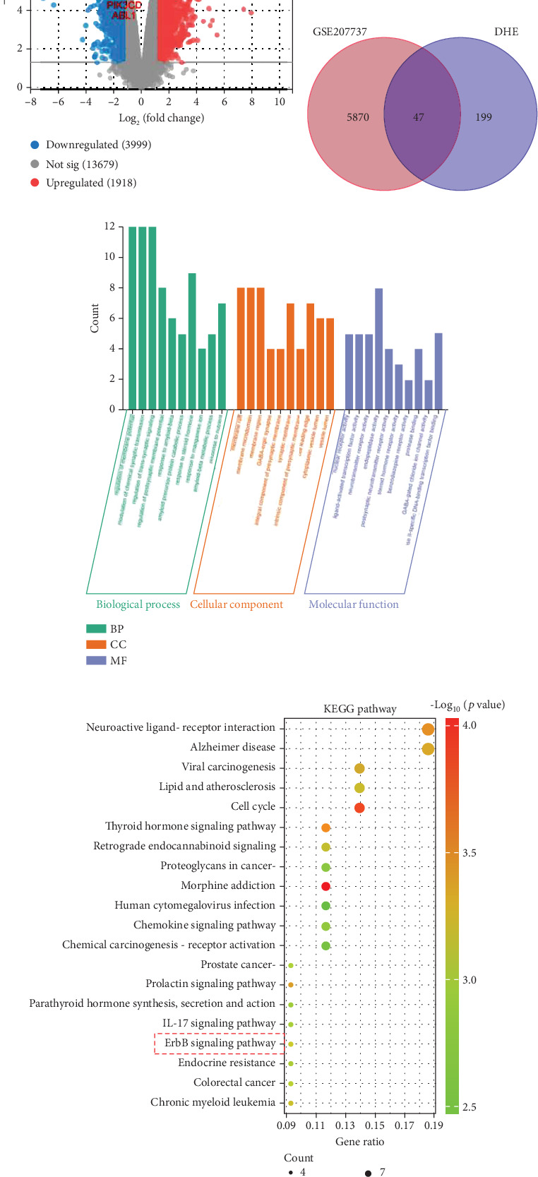
Background: Doxorubicin (DOX) is a widely used antitumor drug; however, its use is limited by the risk of serious cardiotoxicity. Dehydroevodiamine (DHE) is a quinazoline alkaloid which has antiarrhythmic effects. The aim of this study was to investigate the protective effect of DHE on doxorubicin-induced cardiotoxicity (DIC) and its potential mechanism. Materials and Methods: Rat H9c2 cardiomyocytes were exposed to DOX for 24 h to establish a DOX-induced cardiomyocyte injury model. DHE and ErbB inhibitor AG1478 were used to treat H9c2 cells to investigate their effects. Cell counting kit-8 (CCK-8) and lactate dehydrogenase (LDH) release assays were used to evaluate cell viability. Flow cytometry and caspase-3 activity assay were used to detect apoptosis. Western blot was used to detect the expression levels of apoptosis-related proteins and neuregulin-1 (NRG1)/ErbB pathway-related proteins. The levels of proinflammatory cytokines and markers of oxidative stress were also detected, respectively. Quantitative polymerase chain reaction (qPCR) was used to detect mRNA expression levels of hub genes. Results: DHE enhanced cardiomyocyte viability and decreased LDH release in a concentration- and time-dependent manner. DHE also significantly inhibited DOX-induced cardiomyocyte apoptosis, inflammation, and oxidative stress. Bioinformatics analysis showed that the protective mechanism of DHE against DIC was related to ErbB signaling pathway. DOX treatment significantly reduced NRG1, p-ErbB2, and p-ErbB4 protein expression levels in cardiomyocytes, while DHE pretreatment reversed this effect. ErbB inhibitor AG1478 reversed the protective effect of DHE on cardiomyocytes. Conclusion: DHE protects cardiomyocytes against DOX by regulating NRG1/ErbB pathway. DHE may be a potential agent for the prevention and treatment of DIC.

Keywords: NRG1/ErbB pathway; cardiotoxicity; dehydroevodiamine; doxorubicin.


Conflict of interest statement

The authors declare no conflicts of interest.

Figures

Figure 1
Figure 1
DHE enhances the viability of DOX-induced cardiomyocytes. (a) Chemical structure of DHE. (b, c) H9c2 cardiomyocytes were treated with different concentrations of DHE (0, 2.5, 5, 10, 20, and 50 μM) for 24 h, and cell viability was detected by CCK-8 method. (d, e) H9c2 cells were treated with 1 μM DOX and different concentrations (10 and 50 μM) of DHE for 24 h, and then, cell viability and LDH release were measured using CCK-8 and LDH release methods. (f, g) DOX-induced H9c2 cells were treated with 10 μM DHE for different periods (24 or 48 h), and cell viability and LDH release were measured by CCK-8 and LDH assay kit, respectively. All of the experiments were performed in triplicate. ⁣, ⁣∗∗, and ⁣∗∗∗ represent p < 0.05, p < 0.01, and p < 0.001, respectively.
Figure 2
Figure 2
DHE inhibits DOX-induced apoptosis of cardiomyocytes. H9c2 cells were grouped into four groups: control group, 1 μM DOX treatment group, 10 μM DHE treatment group, and 1 μM DOX treatment + 10 μM DHE pretreatment group. (a, b) Flow cytometry was applied to assess apoptosis of H9c2 cells. (c) Caspase-3 activity was detected with caspase-3 detection kit. (d, e) Western blot was used to detect the protein levels of apoptosis-related proteins Bcl-2 and Bax. All of the experiments were performed in triplicate. ⁣∗∗∗ represents p < 0.001.
Figure 3
Figure 3
DHE inhibits DOX-induced inflammation and oxidative stress in cardiomyocytes. H9c2 cells were grouped into four groups: control group, 1 μM DOX treatment group, 10 μM DHE treatment group, and 1 μM DOX treatment + 10 μM DHE pretreatment group. (a–c) The levels of proinflammatory cytokines TNF-α, IL-1β, and IL-6 were detected by ELISA. (d) The intracellular ROS production was detected by DCFH-DA probe. (e–g) The levels of MDA, GSH, and SOD in H9c2 cells were detected. All of the experiments were performed in triplicate. ⁣∗∗ and ⁣∗∗∗ represent p < 0.01 and p < 0.001, respectively.
Figure 4
Figure 4
Functional enrichment analysis of DHE targets in treating DIC. (a) Volcano map for the differentially expressed genes of the GSE207737 dataset. p < 0.05 and |log2 fold change| > 1 are the threshold. Red dots indicate upregulated genes, blue dots downregulated genes, and gray genes with insignificant differences. (b) Venn diagram of DHE-related targets and DIC-related targets. (c) Histogram of GO analysis of DHE targets in DIC treatment. Biological process (BP) is marked by dark cyan, cellular component (CC) is marked by sienna, and molecular function (MF) is marked by steel blue. (d) Bubble map of KEGG pathway enrichment analysis of DHE targets in DIC treatment. The bubble size represents count, and the bubble color represents the p value.
Figure 5
Figure 5
Molecular docking analysis. Molecular docking diagram of DHE with (a) SRC, (b) GSK3B, (c) PIK3CD, (d) ABL1, (e) NRG1, (f) ErbB2, and (g) ErbB4 proteins. Light blue indicates amino acid residues surrounding the binding bag, purple indicates DHE, green indicates macromolecules, and yellow dashed lines indicate hydrogen bonding.
Figure 6
Figure 6
Effect of DHE on the NRG1/ErbB2 pathway in DOX-induced cardiomyocytes. H9c2 cells were grouped into four groups: control group, 1 μM DOX treatment group, 10 μM DHE treatment group, and 1 μM DOX treatment + 10 μM DHE pretreatment group. (a–d) qPCR was used to detect the mRNA expression levels of SRC, GSK3B, PIK3CD, and ABL1. (e, f) Protein expression levels of NRG1, p-ErbB2, and p-ErbB4 were detected by Western blot. All of the experiments were performed in triplicate. ⁣, ⁣∗∗, and ⁣∗∗∗ represent p < 0.05, p < 0.01, and p < 0.001, respectively.
Figure 7
Figure 7
Effects of ErbB inhibitor AG1478 on DOX-induced injury of H9c2 cells after DHE treatment. H9c2 cells were grouped into four groups: control group, 1 μM DOX treatment group, 1 μM DOX treatment + 10 μM DHE pretreatment group, and 1 μM DOX treatment + 10 μM DHE pretreatment + 10 μM AG1478 pretreatment group. (a, b) The cell viability and LDH release of H9c2 cells were measured by CCK-8 assay and LDH release method. (c, d) Apoptosis of H9c2 cells was assessed by flow cytometry. (e) Caspase-3 activity was detected with caspase-3 detection kit. (f–i) qPCR was used to detect the mRNA expression levels of SRC, GSK3B, PIK3CD, and ABL1. (j, k) Western blot was used to detect protein expression levels of Bcl-2, Bax, NRG1, p-ErbB2, and p-ErbB4. All of the experiments were performed in triplicate. ⁣, ⁣∗∗, and ⁣∗∗∗ represent p < 0.05, p < 0.01, and p < 0.001, respectively.
Figure 8
Figure 8
Effects of ErbB inhibition on DOX-induced inflammation and oxidative stress in H9c2 cells after DHE treatment. H9c2 cells were grouped into four groups: control group, 1 μM DOX treatment group, 1 μM DOX treatment + 10 μM DHE pretreatment group, and 1 μM DOX treatment + 10 μM DHE pretreatment + 10 μM AG1478 pretreatment group. (a) The levels of proinflammatory cytokines TNF-α, IL-1β, and IL-6 were detected by ELISA. (b) The intracellular ROS levels were detected by DCFH-DA probe. (c–e) The levels of MDA, GSH, and SOD were detected by the corresponding detection kit. All of the experiments were performed in triplicate. ⁣, ⁣∗∗, and ⁣∗∗∗ represent p < 0.05, p < 0.01, and p < 0.001, respectively.

Similar articles

References

    1. Dozic S., Howden E. J., Bell J. R., Mellor K. M., Delbridge L. M. D., Weeks K. L. Cellular mechanisms mediating exercise-induced protection against cardiotoxic anthracycline cancer therapy. Cells . 2023;12(9):p. 1312. doi: 10.3390/cells12091312. - DOI - PMC - PubMed
    1. Christidi E., Brunham L. R. Regulated cell death pathways in doxorubicin-induced cardiotoxicity. Cell Death & Disease . 2021;12(4):p. 339. doi: 10.1038/s41419-021-03614-x. - DOI - PMC - PubMed
    1. Damiani R. M., Moura D. J., Viau C. M., Caceres R. A., Henriques J. A. P., Saffi J. Pathways of cardiac toxicity: comparison between chemotherapeutic drugs doxorubicin and mitoxantrone. Archives of Toxicology . 2016;90(9):2063–2076. doi: 10.1007/s00204-016-1759-y. - DOI - PubMed
    1. Xiao B., Hong L., Cai X., Mei S., Zhang P., Shao L. The true colors of autophagy in doxorubicin-induced cardiotoxicity. Oncology Letters . 2019;18(3):2165–2172. doi: 10.3892/ol.2019.10576. - DOI - PMC - PubMed
    1. Swain S. M., Whaley F. S., Ewer M. S. Congestive heart failure in patients treated with doxorubicin. Cancer . 2003;97(11):2869–2879. doi: 10.1002/cncr.11407. - DOI - PubMed

MeSH terms

LinkOut - more resources