Multifocal Torpedo Maculopathy Complicated by Choroidal Neovascularization
- PMID: 39742144
- PMCID: PMC11683830
- DOI: 10.1177/24741264241305116
Multifocal Torpedo Maculopathy Complicated by Choroidal Neovascularization
Abstract
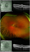
Purpose: To present a pediatric patient with a unique configuration of torpedo maculopathy complicated by macular choroidal neovascularization (CNV). Methods: A single case was retrospectively reviewed. Results: An 8-year-old male child presented with decreased vision in the left eye and was found to have 2 distinct torpedo maculopathy lesions, 1 a smaller hypopigmented lesion in the temporal parafovea and the other a larger hyperpigmented comet-shaped lesion in the temporal periphery. Multimodal imaging showed active CNV. The patient received 2 intravitreal injections of ranibizumab with regression of CNV and recovery of visual acuity. Conclusions: CNV is a rare complication of torpedo maculopathy that can affect pediatric patients in the absence of choroidal excavation. The presence of a hyperpigmented peripheral lesion exhibiting symmetry across the horizontal raphe lends support to the hypothesis that an alteration in the development and migration of retinal pigment epithelium cells across the fetal bulge results in this disorder.
Keywords: anti-VEGF agents; choroidal neovascularization; pediatric retina; retinal pigment epithelium; torpedo maculopathy.
© The Author(s) 2024.
Conflict of interest statement
The authors declared no potential conflicts of interest with respect to the research, authorship, and/or publication of this article.
Figures


Similar articles
-
Multimodal Imaging of Choroidal Structural in Torpedo Maculopathy.Front Med (Lausanne). 2023 Feb 23;10:1085457. doi: 10.3389/fmed.2023.1085457. eCollection 2023. Front Med (Lausanne). 2023. PMID: 36910495 Free PMC article.
-
Multimodal imaging of torpedo maculopathy including adaptive optics.Eur J Ophthalmol. 2020 Mar;30(2):NP27-NP31. doi: 10.1177/1120672119827772. Epub 2019 Feb 8. Eur J Ophthalmol. 2020. PMID: 30732462
-
CHOROIDAL NEOVASCULARIZATION IN TORPEDO MACULOPATHY TREATED BY AFLIBERCEPT: LONG-TERM FOLLOW-UP USING OPTICAL COHERENCE TOMOGRAPHY AND OPTICAL COHERENCE TOMOGRAPHY ANGIOGRAPHY.Retin Cases Brief Rep. 2023 Jul 1;17(4):433-437. doi: 10.1097/ICB.0000000000001213. Retin Cases Brief Rep. 2023. PMID: 37364204
-
A deeper look at torpedo maculopathy.Clin Exp Optom. 2017 Nov;100(6):563-568. doi: 10.1111/cxo.12540. Epub 2017 Apr 23. Clin Exp Optom. 2017. PMID: 28436087 Review.
-
[Polypoidal choroidal vasculopathy].Nippon Ganka Gakkai Zasshi. 2012 Mar;116(3):200-31; discussion 232. doi: 10.4264/numa.71.282. Nippon Ganka Gakkai Zasshi. 2012. PMID: 22568102 Review. Japanese.
Cited by
-
Issues in Medicine: Equity and Site Neutrality.J Vitreoretin Dis. 2025 Mar 16;9(2):125-130. doi: 10.1177/24741264251322832. eCollection 2025 Mar-Apr. J Vitreoretin Dis. 2025. PMID: 40103668 No abstract available.
References
Publication types
LinkOut - more resources
Full Text Sources

