Case Reports
. 2024 Dec 1;16(12):e74913.
doi: 10.7759/cureus.74913. eCollection 2024 Dec.

Medial Opening Wedge Osteotomy for Early Osteoarthritis of the Knee With Dr. Saigal's Plate: A Case Report With Review of Literature

Affiliations
Case Reports

Medial Opening Wedge Osteotomy for Early Osteoarthritis of the Knee With Dr. Saigal's Plate: A Case Report With Review of Literature

Alok C Agrawal et al. Cureus. .

Abstract

Knee pain in patients often involves varus deformity and unicompartmental osteoarthritis (OA). High tibial valgus osteotomy (HTO) is increasingly recognized as an effective treatment, as it realigns the knee's mechanical axis towards the healthier lateral compartment, delaying degenerative changes in the medial compartment and reducing the need for joint replacement. This case report discusses two patients with bilateral knee arthritis and varus deformity who underwent medial opening-wedge high tibial osteotomy (MOWHTO) using Dr. Saigal's plate (Nebula Surgical Pvt. Ltd., Gujarat, India). The first patient, a 45-year-old male with a BMI of 29.3 kg/m², had a good range of motion (ROM) and no ligamentous laxity. The second patient, a 36-year-old female with a BMI of 30 kg/m², also exhibited good knee ROM and no ligamentous laxity. Initial evaluation included comprehensive radiological assessments via four X-rays: anteroposterior (AP) view in 30-degree flexion, lateral view, skyline view for the patellofemoral joint, and a standing orthosonogram view from the hip to the toes. The surgical technique aimed to correct varus angulation with valgus overcorrection. Preoperative preparation followed the Miniaci Method, involving a weight-bearing AP orthoscan of the entire leg to determine the corrective angle. Postoperatively, a protocol focused on fixation rigidity allowed toe-touch walking after six weeks. Suture removal occurred on the 14th day with no NSAIDs administered. Data were collected preoperatively, intraoperatively, and at three, six, and twelve months postoperatively. Primary outcomes included the Oxford Knee Score (OKS), Western Ontario and McMaster Universities Osteoarthritis Index (WOMAC), and ROM. Secondary measures assessed mechanical axis deviation (MAD), correction of varus angulation, pain levels, and complications using the Modified RUST criteria for osteotomy site evaluation. At the final follow-up, both patients showed excellent clinical outcomes with pain-free joint motion and optimal limb alignment. No complications such as infection, hardware failure, or need for total knee replacement were reported. The mean preoperative OKS significantly improved, indicating the procedure's effectiveness in enhancing function and quality of life. The WOMAC pain and functional subscores also improved consistently over the year. Although there was a temporary decrease in knee ROM initially, it rebounded by the final assessments. Overall, the intervention was safe and successful, with no deep infections, deep vein thrombosis, lateral hinge fractures, varus collapse, or implant failures reported.

Keywords: dr saigal; hto; lateral hinge fracture; mechanical axis deviation; medial open wedge osteotomy; medial opening wedge osteotomy; miniaci; modified rust criteria; osteoarthritis (oa); western ontario and mcmaster universities osteoarthritis index.


Conflict of interest statement

Human subjects: Consent for treatment and open access publication was obtained or waived by all participants in this study. Institute Ethical Committee issued approval 2711/IEC-AIIMSRPR/2023. At Institute Ethics Committee meeting held on 21/01/2023,the committee reviewed the research project and the study related document and discussed the ethical issues involved. No ethical issues was identified.Hence ,IEC deided to approve the above referenced project. . Conflicts of interest: In compliance with the ICMJE uniform disclosure form, all authors declare the following: Payment/services info: All authors have declared that no financial support was received from any organization for the submitted work. Financial relationships: All authors have declared that they have no financial relationships at present or within the previous three years with any organizations that might have an interest in the submitted work. Other relationships: All authors have declared that there are no other relationships or activities that could appear to have influenced the submitted work.

Figures

Figure 1
Figure 1. A) Pre-operative X-ray in weight bearing anteroposterior and lateral views. B) Orthoscannogram of both lower limbs in weight bearing standing position showing varus angulation.
Figure 2
Figure 2. A) Pre-operative clinical images showing mild Varus deformity at both knee joints. B) Clinical image showing preoperative knee range of flexion.
Figure 3
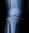
Figure 3. Illustrative diagram showing the calculation of the Alpha angle using the Miniaci method. A) To establish Mikulicz’s mechanical axis, draw a line from the femoral head to the ankle joint (α) and mark the new ankle center (b). Identify the hinge point (h) at the upper tibial metaphysis, then connect points (h) to (a) and rotate to intersect (b). Finally, connect (b) and (h) to form the angle α. B) This correction angle is translated at the medial osteotomy site 4 cm below the medial joint line extending up to lateral hinge.
Figure 4
Figure 4. Identification of lateral hinge point.
Hinge point is marked with K- Wires which is entered immediately superior to the pes-anserinus tendons. The osteotomy's hinge is located in the upper 1/3rd of the tibio-fibular joint—Medial Wedge Opening HTO surgical Technique
Figure 5
Figure 5. Implant characteristics of Saigal's plate.
Figure 6
Figure 6. A) Minimally invasive approach to proximal medial tibia for MOWHTO. B) Subperiosteal dissection up to the proximal medial tibia.
Figure 7
Figure 7. A, B) 2 Parallel K-wires are inserted aiming towards the lateral hinge point to mark the site of osteotomy. C) Identification and marking of the lateral hinge point using K wires.
Figure 8
Figure 8. A) Horizontal osteotomy is done using the K wire as a guide. B) Spreading the osteotomy site using multiple chisels.
Figure 9
Figure 9. A, B) Fixation of Saigal's plate to protect the osteotomy site.
Figure 10
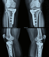
Figure 10. A) Immediate post-operative X-ray showing Implant in good position and the deformity was corrected to acceptable range. B) Three months follow-up X-ray showing remodeling at the osteotomy site.
Figure 11
Figure 11. A) Complete extension of the right knee up to 0 degrees at one year of follow-up. B) Flexion more than 90 degrees in the right knee at one year of follow-up. C) Extension at left knee up to 0 degrees at one year of follow-up. D) Flexion more than 90 degrees in the left knee at one year of follow-up.
Figure 12
Figure 12. One-year follow-up X-ray showing healing of the osteotomy site with intact lateral hinge.
Figure 13
Figure 13. Preoperative clinical images. A) Standing weight-bearing front view. B) Maximum flexion of right knee. C) Maximum flexion of left knee joint. D) Lower limb alignment in supine position.
Figure 14
Figure 14. A) Radiograph showing anteroposterior view of the knee joint in standing weight bearing position. B, C) Radiograph showing lateral view of both knees. D) Standing orthoscannogram of both lower limb in weight bearing posture.
Figure 15
Figure 15. A) Immediate post-operative radiograph of MOWHTO using Dr Saigal's plate. B) Six months of follow-up X-ray showing progress of union in both knees.
Figure 16
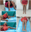
Figure 16. A) Knee flexion on immediate post-operative day 5. B, C) Knee flexion of Right and left knee respectively at one year of follow-up. D) Standing weight bearing clinical image at one-year of follow-up. E) Lower limb alignment at one-year of follow-up.
Figure 17
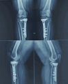
Figure 17. Anteroposterior and lateral radiographs at one year of follow-up showing union of all the cortices. The fracture line is visible only in the AP view of the right knee.

References

    1. Tibial osteotomy for osteoarthritis of the knee. JA JP, WA W. J Bone Joint Surg Br. 1961;43-B:746–751. - PubMed
    1. High tibial osteotomy using a locking titanium plate with or without autografting. Sarman H, Isik C, Uslu M, Inanmaz ME. Acta Ortop Bras. 2019;27:80–84. - PMC - PubMed
    1. The short-term follow-up results of open wedge high tibial osteotomy with using an Aescula open wedge plate and an allogenic bone graft: the minimum 1-year follow-up results. Lee SC, Jung KA, Nam CH, Jung SH, Hwang SH. Clin Orthop Surg. 2010;2:47–54. - PMC - PubMed
    1. Comparison between autogenous bone graft and allogenous cancellous bone graft in medial open wedge high tibial osteotomy with 2-year follow-up. Cho SW, Kim DH, Lee GC, Lee SH, Park SH. Knee Surg Relat Res. 2013;25:117–125. - PMC - PubMed
    1. Improvements in surgical technique of valgus high tibial osteotomy. Lobenhoffer P, Agneskirchner JD. Knee Surg Sports Traumatol Arthrosc. 2003;11:132–138. - PubMed

Publication types

LinkOut - more resources