

Trigeminal Nerve Schwannoma: A Rare Case of Multisymptomatic Cranial Nerve Involvement
- PMID: 39742186
- PMCID: PMC11687709
- DOI: 10.7759/cureus.74919
Trigeminal Nerve Schwannoma: A Rare Case of Multisymptomatic Cranial Nerve Involvement
Abstract
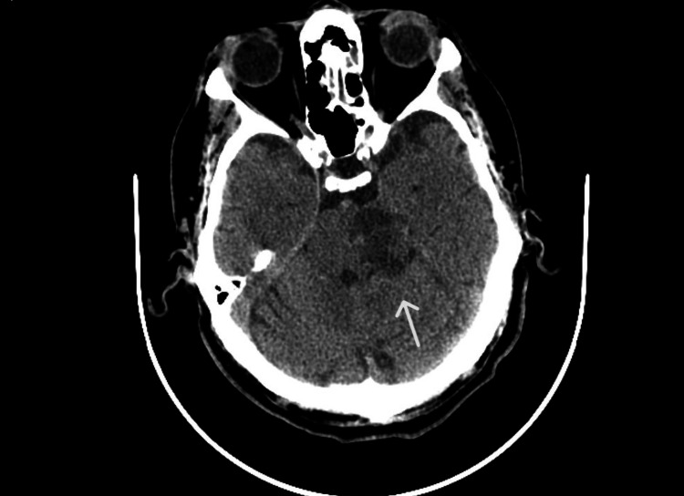
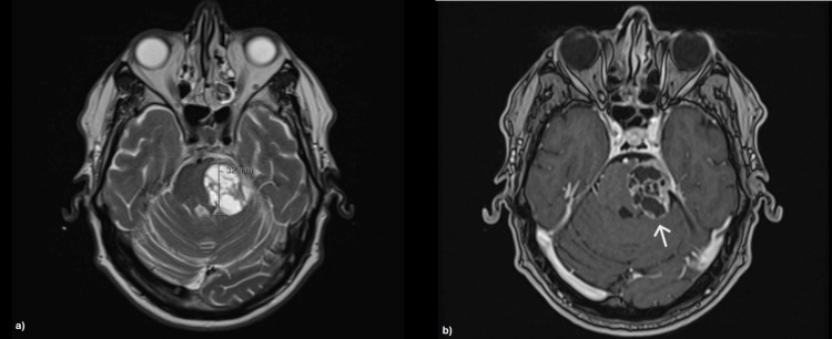
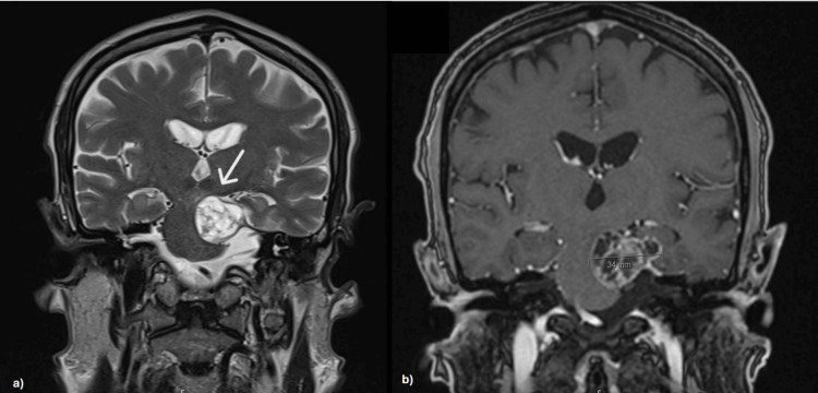
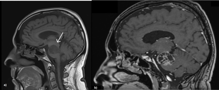
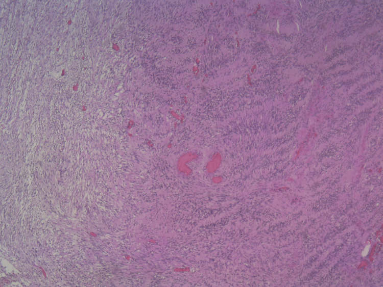
Schwannomas (SCs) are benign tumors composed of neoplastic Schwann cells and are relatively uncommon intracranially. Although these tumors are frequently associated with neurofibromatosis type 2 (NF2), they may also arise idiopathically, and their pathogenesis remains poorly understood. A 70-year-old Caucasian man presented with a two-month history of vertigo, gait imbalance, and decreased visual acuity in the left eye accompanied by photophobia, nausea, vomiting, and occasional headaches. On physical examination, he exhibited hyposmia, horizontal nystagmus, superior oblique palsy, decreased photoreactivity of the left pupil, hypoesthesia in the middle and lower left facial regions, and a positive Romberg sign. Non-contrast computed tomography (CT) revealed a hypodense, expansive lesion in the left mesencephalic-pontine region. Based on the radiological characteristics, the radiologist suggested ischemic injury or neoplasm as the leading diagnostic hypothesis. Magnetic resonance imaging (MRI) with contrast revealed a cystic-necrotic lesion with multiloculated characteristics in the left mesencephalic-pontine area, with significant mass effect and compression of the brainstem and adjacent ventricular pathway. The lesion was suspected to be a schwannoma, and the patient underwent surgical resection via left temporal and suboccipital craniotomy. Histopathological examination confirmed a schwannoma of the left trigeminal nerve. SCs are most commonly diagnosed in the second and third decades of life, often growing slowly and remaining asymptomatic until they reach a size sufficient to cause functional impairment. MRI is the imaging modality of choice, but histologic confirmation remains the gold standard for diagnosis. This case underscores the rarity of trigeminal nerve SCs presenting with symptoms resulting not only from the affected nerve but also from compression of adjacent structures, such as the vestibulocochlear and abducens nerves. It also highlights the importance of maintaining a high index of suspicion when diagnosing rare intracranial SCs. Advanced imaging techniques and comprehensive clinical evaluation are crucial for identifying complex neurological conditions, particularly when initial findings, such as CT results, suggest alternative diagnoses like stroke.
Keywords: gait imbalance; mesencephalic-pontine lesion; neurological examination; nystagmus; schwannoma; trigeminal nerve.
Copyright © 2024, Pinheiro et al.
Conflict of interest statement
Human subjects: Consent for treatment and open access publication was obtained or waived by all participants in this study. Conflicts of interest: In compliance with the ICMJE uniform disclosure form, all authors declare the following: Payment/services info: All authors have declared that no financial support was received from any organization for the submitted work. Financial relationships: All authors have declared that they have no financial relationships at present or within the previous three years with any organizations that might have an interest in the submitted work. Other relationships: All authors have declared that there are no other relationships or activities that could appear to have influenced the submitted work.
Figures
Similar articles
-
Cystic Progression of a Cavernous Malformation at the Level of the Trigeminal Root Entry Zone Presenting With Sudden Onset of Trigeminal Neuralgia.J Craniofac Surg. 2018 Nov;29(8):e728-e730. doi: 10.1097/SCS.0000000000004501. J Craniofac Surg. 2018. PMID: 29570519
-
Diagnostic Value of Preoperative Electrodiagnostic Analysis in a Patient with Facial Palsy and a Large Vestibular Schwannoma: Case Report.Diagnostics (Basel). 2022 Feb 20;12(2):542. doi: 10.3390/diagnostics12020542. Diagnostics (Basel). 2022. PMID: 35204631 Free PMC article.
-
Diagnosis and management of bilateral vestibular schwannoma in the cerebellopontine angle: A rare case report.Radiol Case Rep. 2024 Jan 13;19(4):1271-1275. doi: 10.1016/j.radcr.2023.12.042. eCollection 2024 Apr. Radiol Case Rep. 2024. PMID: 38292801 Free PMC article.
-
Pediatric intracranial lower cranial nerve schwannoma unassociated with neurofibromatosis type 2: case report and review of the literature.Childs Nerv Syst. 2019 Jun;35(6):1041-1044. doi: 10.1007/s00381-018-04045-4. Epub 2019 Jan 13. Childs Nerv Syst. 2019. PMID: 30637480 Review.
-
Abducens nerve schwannoma: a case report and review of the literature.Surg Neurol. 2002 Mar;57(3):183-8; discussion 188-9. doi: 10.1016/s0090-3019(01)00670-x. Surg Neurol. 2002. PMID: 12009546 Review.
References
-
- Schwannoma of the 6th nerve: case report and review of the literature. Li X, Li J, Li J, Wu Z. Chin Neurosurg J. 2015;1:5.
Publication types
LinkOut - more resources
Full Text Sources
Research Materials
Miscellaneous