. 2025 Jan 3;11(1):eado6894.
doi: 10.1126/sciadv.ado6894. Epub 2025 Jan 1.

Blood-derived APLP1+ extracellular vesicles are potential biomarkers for the early diagnosis of brain diseases

Affiliations

Blood-derived APLP1+ extracellular vesicles are potential biomarkers for the early diagnosis of brain diseases

Yuri Choi et al. Sci Adv. .

Abstract

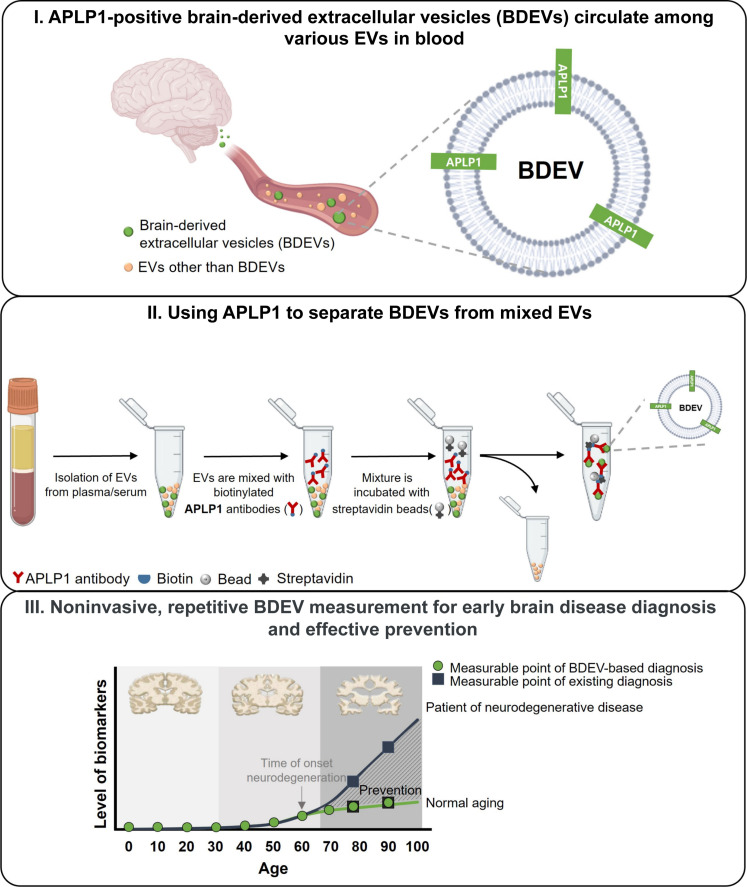
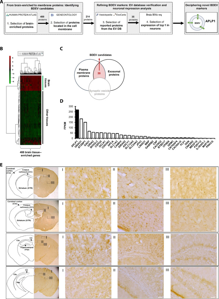
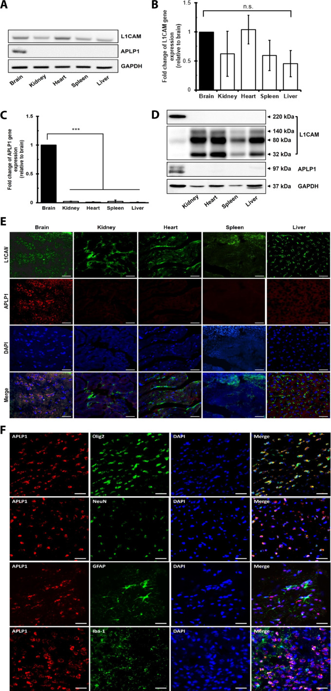
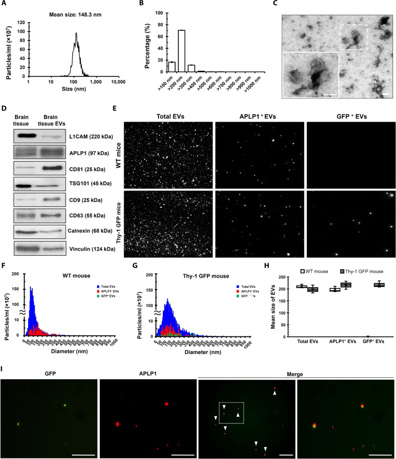
The early detection of neurodegenerative diseases necessitates the identification of specific brain-derived biomolecules in peripheral blood. In this context, our investigation delineates the role of amyloid precursor-like protein 1 (APLP1)-a protein predominantly localized in oligodendrocytes and neurons-as a previously unidentified biomarker in extracellular vesicles (EVs). Through rigorous analysis, APLP1+ EVs from human sera were unequivocally determined to be of cerebral origin. This assertion was corroborated by distinctive small RNA expression patterns of APLP1+ EVs. The miRNAs' putative targets within these EVs manifested pronounced expression in the brain, fortifying their neurospecific provenance. We subjected our findings to stringent validation using Thy-1 GFP M line mice, transgenic models wherein GFP expression is confined to hippocampal neurons. An amalgamation of these results with an exhaustive data analysis accentuates the potential of APLP1+ EVs as cerebrally originated biomarkers. Synthesizing our findings, APLP1+ EVs are postulated not merely as diagnostic markers but as seminal entities shaping the future trajectory of neurodegenerative disease diagnostics.


Figures

Fig. 1.
Fig. 1.. A schematic representation elucidating the diagnostic potential of APLP1+ BDEVs in blood for neurodegenerative conditions.
The bloodstream contains a mixture of EVs: those derived from the brain (BDEVs, depicted as small green circles) and others originating from various organs (represented as small light orange circles). A distinguishing feature of BDEVs is the presence of the brain-specific protein APLP1 on their membrane (step I). Leveraging an APLP1-specific antibody, it becomes feasible to selectively isolate these BDEVs from the milieu of circulating EVs, filtering out vesicles released from other organs (step II). Periodic assessments leveraging this BDEV-centric approach can facilitate early disease detection, allowing for real-time monitoring of brain health. Such proactive tracking has the potential to preempt or mitigate disease progression, offering another avenue in neurodegenerative disease management (step III).
Fig. 2.
Fig. 2.. Comprehensive approach for BDEV biomarker identification.
(A) An in-depth in silico workflow delineating the process for unearthing brain-specific BDEV markers. (B) A heatmap detailing the RNA expression for brain-specific proteins across different human tissues, providing a visual representation of tissue-specific expression. (C) A Venn diagram illustrating the convergence of our identified brain-specific plasma membrane proteins with entries from the ExoCarta and Vesiclepedia databases, resulting in 35 potential BDEV markers. (D) A comparative analysis of the expression magnitudes of the 35 BDEV candidates based on their FPKM (fragments per kilobase per million reads) values in neurons, drawing data from the Brain RNA-seq database. (E) Immunohistochemical panels from seven independent assays depict DAB staining localized to APLP1 within diverse cerebral territories. Brain sections derived from seven C57BL/6 mice. The images on the left provide an overarching perspective of the whole brain sections, emphasizing the pervasive presence of APLP1 (×2 magnification). Images on the right (I to III) are enlarged (×200 magnification) highlighting APLP1 distribution across specific brain areas. Additional data can be explored in fig. S1. Scale bars, 50 μm. Ctx, cerebral cortex; Str, striatum; CC, corpus callosum; Hip, hippocampus.
Fig. 3.
Fig. 3.. Brain-specific expression of APLP1.
(A) mRNA profiles of APLP1 and L1CAM across different mouse organs. (B and C) Relative mRNA expression levels quantified via reverse transcription qPCR, normalized to glyceraldehyde phosphate dehydrogenase (GAPDH). Data are presented as the means ± SEM of eight independent experiments, and statistical analysis was performed on 2ΔCt values using the analysis of variance (ANOVA; post hoc: Tukey). Symbol “***” indicates a value of < 0.001, respectively, compared with the brain. (D and E) Western blot representation (D) and immunofluorescence imaging (E) of L1CAM and APLP1 across varied tissues derived from five C57BL/6 mice. (F) Use of RNAscope and IHC to detect APLP1 colocalization in mouse cerebral cortex cells. The mRNA of APLP1 is visualized in red, while oligodendrocytes (Olig2), neurons (NeuN), astrocytes (GFAP), or microglial cells (Iba-1) are visualized in green fluorescence. Scale bars, 50 μm. The experiment was performed with three C57BL/6 mice. n.s., not significant; DAPI, 4′,6-diamidino-2-phenylindole.
Fig. 4.
Fig. 4.. APLP1+ EVs originate from the brain.
(A) NTA-derived graphs showing size and concentration metrics of the brain tissue EVs. (B) Percentage-based size distributions of EVs, derived from the data presented in (A). (C) TEM images, at ×10,000 and ×20,000 magnifications, depicting EV morphology. Scale bars, 100 nm. Error bars in the figures delineate the SE of the mean (±SEM) from triplicate measurements of brain tissue EVs. (D) Western blot of APLP1 and EV markers in brain tissues and brain tissue EVs from five C57BL/6 mice. (E) ZetaView analysis of plasma-derived EVs in WT and Thy-1 GFP M line mice. Total EVs appear in light-scatter mode, GFP+ EVs in 488-nm fluorescence mode, and APLP1+ EVs in 640-nm fluorescence mode. (F and G) EV size distribution in WT and Thy-1 GFP M line mice using ZetaView. (H) Average EV size ranges between 198 and 217.8 nm in both mouse types. (I) Immunostaining of APLP1 in plasma EVs from Thy-1 GFP M line mice, with white arrowheads highlighting APLP1+GFP+ EVs. Scale bars, 25 μm. All data were obtained from at least three mice.
Fig. 5.
Fig. 5.. Comprehensive characterization of APLP1-enriched EVs from human plasma.
(A) TEM images showing the morphology of mouse plasma EVs, human plasma EVs, and APLP1+ EVs. Scale bars, 100 nm. (B) Representative florescence staining images. Overlay of APLP1-stained EVs (in red) and brain cell markers (in green) from human plasma. Scale bars, 10 μm. (C) Size distribution patterns of various EV samples determined via NTA. (D) Modal size comparisons of EV populations. (E) Concentrations of EVs, highlighting the relative abundance of APLP1+ EVs in the overall plasma EV pool. Data are presented as means ± SEM. (F) A comparative analysis of mRNA expression levels for neuronal markers in human neuronal cell-derived EVs (hNDEVs), APLP1+ EVs, and the residual EVs. (G) Western blot showing the neuronal markers in APLP1+ EVs compared with those in residual EVs from human plasma. Results were based on at least three separate experiments. NSE, neuron-specific enolase.
Fig. 6.
Fig. 6.. Comprehensive analysis of protein and miRNA profiles in APLP1+ EVs and their neurological implications.
(A) Venn diagram showing the overlap of proteins identified in both brain organoid–derived EVs and APLP1+ EVs. (B) Bar chart delineating the GO term analysis of proteins detected in both the brain organoid–derived EVs and APLP1+ EVs, with a specific emphasis on brain-associated GO terms. The y axis enumerates distinct GO terms, while the x axis indicates the −log10 adjusted P value. This visualization offers insights into brain-functional annotations derived from the GO enrichment analysis. (C) Heatmap illustrating the differential expression patterns of miRNAs between APLP1+ EVs and residual EVs. Within the heatmap, rows have been centered, and unit variance scaling is applied to the normalized expression data. Shades of red denote elevated expression levels, whereas green shades indicate reduced expression levels. The Pos indicate APLP+ EVs and the Neg indicate Residual EVs. (D) Principal components analysis (PCA) visualizing the variance in miRNA profiles between APLP1+ EVs and residual EVs. (E) Volcano plot that elucidates the differential miRNA expression between APLP1+ EVs and residual EVs. The graph’s orientation allows for easy identification of miRNAs that are either more abundant in APLP1+ EVs (points to the right) or residual EVs (points to the left). The y axis offers a perspective on the −log10 adjusted P value, providing a quick gauge of significance. (F) A GO term analysis on the targets of up-regulated miRNAs in APLP1+ EVs, leveraging the miEAA tool. Brain-associated terms are highlighted, with nine terms significantly enriched in APLP1+ EVs, in stark contrast to the absence of such terms in residual EVs. The EVs used in the analysis were obtained from plasma of six volunteers.
Fig. 7.
Fig. 7.. Diagnostic potential of APLP1+ EVs in neurological disorders.
(A) Enzyme-linked immunosorbent assay (ELISA)–derived absorbance units representing the levels of EV markers (CD9, CD81, and CD63) and key antigens (EGFR, EGFRviii, L1CAM, and APLP1) within plasma EVs from the healthy group and the GBM patient group. Data represent means ± SEM of three independent experiments. (B) Representative immunostaining images contrasting plasma EVs from healthy individuals and patients with GBM (n = 3). The white arrowheads indicates CD63+APLP1+ EVs. Scale bars, 5 μm. (C) Quantification of CD63+APLP1 EVs and CD63+APLP1+ EVs in each group (n = 3). (D) Fold change ratio of CD63+APLP1 EVs and CD63+APLP1+ EVs between the healthy and GBM groups (n = 3). Data represent means ± SEM of three independent experiments. Statistical analysis was conducted using the ANOVA test (post hoc: Tukey). **P < 0.01 and ***P < 0.001, statistical differences.

Similar articles

Cited by

References

    1. Katsuno M., Sahashi K., Iguchi Y., Hashizume A., Preclinical progression of neurodegenerative diseases. Nagoya J. Med. Sci. 80, 289–298 (2018). - PMC - PubMed
    1. Lee J. C., Kim S. J., Hong S., Kim Y., Diagnosis of Alzheimer’s disease utilizing amyloid and tau as fluid biomarkers. Exp. Mol. Med. 51, 1–10 (2019). - PMC - PubMed
    1. de Aquino C. H., Methodological issues in randomized clinical trials for prodromal Alzheimer’s and Parkinson’s disease. Front. Neurol. 12, 694329 (2021). - PMC - PubMed
    1. Ricci M., Cimini A., Chiaravalloti A., Filippi L., Schillaci O., Positron emission tomography (PET) and neuroimaging in the personalized approach to neurodegenerative causes of dementia. Int. J. Mol. Sci. 21, 7481 (2020). - PMC - PubMed
    1. Marquez F., Yassa M. A., Neuroimaging biomarkers for Alzheimer’s disease. Mol. Neurodegener. 14, 21 (2019). - PMC - PubMed