. 2025 Jan;17(779):eadi6148.
doi: 10.1126/scitranslmed.adi6148. Epub 2025 Jan 1.

G6PC2 controls glucagon secretion by defining the set point for glucose in pancreatic α cells

Affiliations

G6PC2 controls glucagon secretion by defining the set point for glucose in pancreatic α cells

Varun Bahl et al. Sci Transl Med. 2025 Jan.

Abstract

Elevated glucagon concentrations have been reported in patients with type 2 diabetes (T2D). A critical role for α cell-intrinsic mechanisms in regulating glucagon secretion was previously established through genetic manipulation of the glycolytic enzyme glucokinase (GCK) in mice. Genetic variation at the glucose-6-phosphatase catalytic subunit 2 (G6PC2) locus, encoding an enzyme that opposes GCK, has been reproducibly associated with fasting blood glucose and hemoglobin A1c. Here, we found that trait-associated variants in the G6PC2 promoter are located in open chromatin not just in β but also in α cells and documented allele-specific G6PC2 expression of linked variants in human α cells. Using α cell-specific gene ablation of G6pc2 in mice, we showed that this gene plays a critical role in controlling glucose suppression of amino acid-stimulated glucagon secretion independent of alterations in insulin output, islet hormone content, or islet morphology, findings that we confirmed in primary human α cells. Collectively, our data demonstrate that G6PC2 affects glycemic control via its action in α cells and possibly suggest that G6PC2 inhibitors might help control blood glucose through a bihormonal mechanism.


Conflict of interest statement

Competing interests: The authors declare that they have no competing interests, no consulting agreements and that no patent has been filed regarding the data presented in this study.

Figures

Fig. 1:
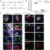
Fig. 1:. The islet-specific glucose-6-phosphatase catalytic subunit G6PC2 is expressed in human α cells.
(A) Snapshot of the G6PC2 region from the UCSC Genome Browser (build hg19) showing the following tracks: reliable α and β cell peaks from bulkATAC-seq; snATAC-seq signal for α, β, and acinar cells; and fasting plasma glucose (FPG)-relevant SNPs. SNPs in high LD with each other (based on r2 in EUR) are shown in the same color. (B) Immunostaining of human non-diabetic pancreatic sections for G6PC2 showing G6PC2 immunoreactivity in a subset of α cells. Scale bar equals 10 μm. (C) Allele-specific read count percentages in α cells from deceased European ancestry organ donors heterozygous for the reference (C) and alternate (G) alleles of rs2232328 from bulk RNA-seq data. P-values are from one-sided binomial tests on the read counts: *p<0.05, **p<0.01. Donor labels are colored by disease status group: black=control, orange=non diabetic auto-antibody positive, red=T2D.
Fig. 2:
Fig. 2:. Efficient ablation of G6pc2 in Gcg+ cells in G6pc2loxP/loxP; Gcg-CreERT2 mice.
(A) Design of conditional null mutation in G6pc2: (top) G6pc2 gene structure, with triangles indicating binding sites for gRNAs used for Cas9-assisted gene targeting, (middle) repair templates for introduction of the 5’ and 3’ loxP sites, where loxP sites (blue arrowhead) are marked by SalI or SpeI restriction endonuclease sites, and (bottom) targeted conditional null allele, with PCR primers used for genotyping (arrows). (B) Ablation efficiency of G6pc2 from glucagon (GCG)-positive cells in the α-G6PC2KO model. N = 5. P-value from unpaired Student’s t-test: ****p<0.0001. (C) Representative immunofluorescent staining of G6pc2 and glucagon in the pancreas from α-G6PC2KO and control mice. (D) Immunofluorescent staining of dispersed islet cells obtained from α-G6PC2KO and control mice for G6PC2, glucagon, and insulin (INS) demonstrating the presence of G6PC2 in both α and β cells from control mice but only in β cells from α-G6PC2KO mice. Yellow arrows point to G6PC2-positive α cells, Green arrows point to G6PC2-negative α cells. P-value from unpaired Student’s t-test: ****p<0.0001.
Fig. 3:
Fig. 3:. Increased 2-NBDG retention in islets from α-G6PC2KO mice and G6PC2-ablated human α cells.
Isolated mouse pancreatic islets were incubated with 2-NBDG for 30 minutes, and 2-NBDG fluorescence was measured at (A) 0 minutes, (B) 5 minutes, and (C) 15 minutes post-washout with glucose-free media. (n=5 mice for α-G6PC2KO, n=11 mice total for G6pc2loxP/loxP and Gcg-CreERT2). (D) Human α cells transduced with shRNA against G6PC2 or non-silencing (NS) control were incubated with 2-NBDG for 40 min followed by serial images taken post-washout at time zero and in 10 minutes intervals to measure the rate of 2-NBDG efflux. Green – 2NBDG, blue - DNA. (E) Decay curves representing the mean 2-NBDG signal from single α cells presented as % of basline, ± SE. (F) Bars represent the median rate of 2-NBDG efflux of 42 shG6PC2 and 44 NS α cells, respectively. P-values from unpaired Student’s t-test: ****p<0.0001.
Fig. 4:
Fig. 4:. α cell-specific G6PC2 ablation alters glucose homeostasis in adult male mice.
(A) Body weights of male α-G6PC2KO and control mice (n=9 for α-G6PC2KO, n=11 total for G6pc2loxP/loxP and Gcg-CreERT2). (B) Ad libitum blood glucose for male α-G6PC2KO and control mice (n=9 for α-G6PC2KO, n=11 total for G6pc2loxP/loxP and Gcg-CreERT2). (C) Intraperitoneal glucose tolerance test (1mg/g bodyweight) (n=9 for α-G6PC2KO, n=11 total for G6pc2loxP/loxP and Gcg-CreERT2). (D) Area Under the Curve (AUC) from intraperitoneal glucose tolerance test in (C). (E) Plasma glucagon and (F) plasma insulin in mice fasted for 16 hours or 5 min after glucose injection in (C). (G) 16 hour fasted blood glucose values (n=9 for α-G6PC2KO, n=11 total for G6pc2loxP/loxP and Gcg-CreERT2). (H) Intraperitoneal insulin tolerance test (0.75U/kg bodyweight) (n=9 for α-G6PC2KO, n=11 total for G6pc2loxP/loxP and Gcg-CreERT2). (I) Area Under the Curve between 30- and 90-min post-insulin injection (AUC30–90) from intraperitoneal insulin tolerance test in (H). (J) Plasma glucagon in mice fasted for 4 hours or 30 min after insulin injection in (H). (*p < 0.05, **p < 0.01, ***p < 0.001 vs. genetically unmodified control). P-values from unpaired Student’s t-test or two-way ANOVA with post hoc Bonferroni test: *p < 0.05, **p < 0.01, ***p < 0.001. TMX, Tamoxifen.
Fig. 5:
Fig. 5:. Ablation of α cell G6PC2 impairs the counter-regulatory response to phloridzin-induced hypoglycemia.
(A) Ad libitum blood glucose in female α-G6PC2KO and control mice (n=8 for control vehicle, n=4 for α-G6PC2KO vehicle, n=8 for control phloridzin, n=6 for α-G6PC2KO phloridzin) monitored on days 0, 7, and 14. (B) Ad libitum blood glucose for phloridzin-treated α-G6PC2KO and control mice on days 7 and 14 in (A) (n=5 for control phloridzin, n=4 for α-G6PC2KO phloridzin). (C) Plasma glucagon and (D) insulin were assessed on days 0, 7, and 14 for the phloridzin-induced hypoglycemia experiment in (A). P-values from unpaired Student’s t-test or two-way ANOVA with post hoc Bonferroni test: *p<0.05.
Fig. 6:
Fig. 6:. Glucose is more potent in suppressing glucagon secretion in isolated pancreatic islets from male α-G6PC2KO mice.
(A) Glucagon secretion and (B) content from isolated pancreatic islets in the presence of the indicated glucose concentrations with 4mM amino acid mixture or 30mM KCl (n=9 for α-G6PC2KO, n=11 total for G6pc2loxP/loxP and Gcg-CreERT2). (C) Insulin secretion and (D) content from isolated pancreatic islets in the presence of the indicated glucose concentrations with 4mM amino acid mixture or 30mM KCl (n=9 for α-G6PC2KO, n=11 total for G6pc2loxP/loxP and Gcg-CreERT2). P-values from two-way ANOVA with post hoc Bonferroni test:*p < 0.05, ****p < 0.0001.
Fig. 7:
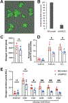
Fig. 7:. shRNA-mediated ablation of G6PC2 limits glucagon release in α cell-enriched human pseudo-islets.
(A) Human α cell pseudo-islets transduced with lentiviral particles expressing GFP and shRNA against G6PC2 (shG6PC2), 20X magnification. (B) Bars represent remaining expression of G6PC2 after transduction of human α cells with lentivirus carrying shRNA against G6PC2 or non-silencing control, determined by qPCR. n=3 human donors. (C) Glucagon content. Total glucaon content of the pseudo-islets was determined after the end of the experiment were quantified using ELISA. (D) Physiological amino acid (AA) mixture and depolarization with KCl stimulated glucagon release from α cell pseudoislets. (E) Glucagon secretion in pseudo-islets transduced with lentiviral particles expressing shRNA against G6PC2 and GFP (G6PC2, blue) or a non-silencing control (NS, red), 12 pseudo-islets per replicate, 4–5 replicates per condition. Represenative data from one of four independent studies with similar outcomes shown. P-values was determined by unpaired Student’s t-test. *, p< 0.05, **, p<0.01, ***, p<0.001, NS, not-significant.

References

    1. Gerich JE, Charles MA, Grodsky GM, Regulation of pancreatic insulin and glucagon secretion. Annu Rev Physiol 38, 353–388 (1976). - PubMed
    1. Quesada I, Tuduri E, Ripoll C, Nadal A, Physiology of the pancreatic alpha-cell and glucagon secretion: role in glucose homeostasis and diabetes. J Endocrinol 199, 5–19 (2008). - PubMed
    1. Zhang P, Zhang X, Brown J, Vistisen D, Sicree R, Shaw J, Nichols G, Global healthcare expenditure on diabetes for 2010 and 2030. Diabetes Res Clin Pract 87, 293–301 (2010). - PubMed
    1. Unger RH, Orci L, The essential role of glucagon in the pathogenesis of diabetes mellitus. Lancet 1, 14–16 (1975). - PubMed
    1. D'Alessio D, The role of dysregulated glucagon secretion in type 2 diabetes. Diabetes Obes Metab 13 Suppl 1, 126–132 (2011). - PubMed

LinkOut - more resources