Review
. 2024 Dec 31;38(4):309-325.
doi: 10.4285/ctr.24.0058.

Targeting T helper 17 cells: emerging strategies for overcoming transplant rejection

Affiliations
Review

Targeting T helper 17 cells: emerging strategies for overcoming transplant rejection

Young Joon Lee et al. Clin Transplant Res. .

Abstract

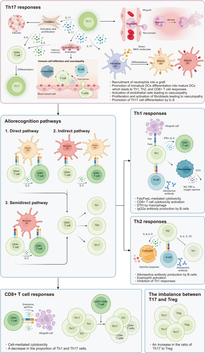
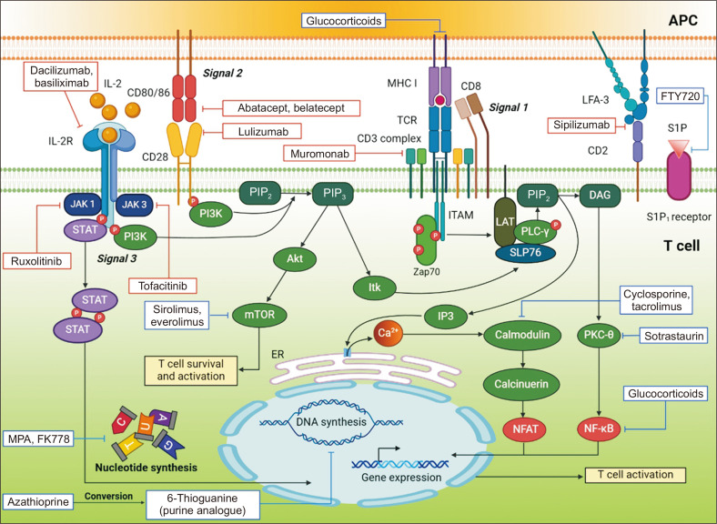
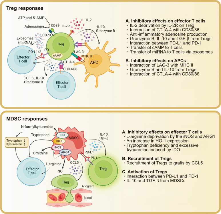
Solid organ transplantation has significantly improved the survival rate of patients with terminal organ failure. However, its success is often compromised by allograft rejection, a process in which T helper 17 (Th17) cells play a crucial role. These cells facilitate rejection by enhancing neutrophil infiltration into the graft and by activating endothelial cells and fibroblasts. Additionally, Th17 cells can trigger the activation of other T cell types, including Th1, Th2, and CD8+ T cells, further contributing to rejection. An imbalance between Th17 and regulatory T cells (Tregs) is known to promote rejection. To counteract this, immunosuppressive drugs have been developed to inhibit T cell activity and foster transplant tolerance. Another approach involves the adoptive transfer of regulatory cells, such as Tregs and myeloid-derived suppressor cells, to dampen T cell functions. This review primarily focuses on the roles of Th17 cells in rejection and their interactions with other T cell subsets. We also explore various strategies aimed at suppressing T cells to induce tolerance.

Keywords: Allograft rejection; Immune tolerance; Th17 cells; Transplantation.


Conflict of interest statement

Conflict of Interest

No potential conflict of interest relevant to this article was reported.

Figures

Fig. 1
Fig. 1
Roles of T cell subtypes in allograft rejection. Th17 cells promote the infiltration of neutrophils into a graft in an interleukin (IL)-17A-independent pathway [9]. Neutrophils undergo NETosis to promote allograft rejection [10,11]. Th17 cells also activate endothelial cells and fibroblasts. Endothelial cells release chemokines and increase their expression of adhesion molecules. In turn, immune cells are recruited and move from the bloodstream across the endothelial monolayer into the blood vessel wall. This immune cell infiltrate is a hallmark of transplant vasculopathy [12]. Activated fibroblasts contribute to fibrosis [13]. Activated endothelial cells and fibroblasts secrete IL-6, promoting the differentiation of Th17 cells [14]. IL-17 modulates allograft rejection by promoting the maturation of dendritic cells (DCs) [15]. Compared to immature DCs, which are specialized in endocytosis, mature DCs express higher levels of MHC and costimulatory molecules on their surface for efficient antigen presentation [16]. Mature DCs present peptides with MHC molecules to activate CD4+ and CD8+ T cells. The peptides are produced by the processing of donor MHC molecules. CD8+ and CD4+ T cells recognize the peptides via the direct, indirect, and semidirect pathways. In the direct pathway, recipient CD8+ T cells and CD4+ T cells engage complexes of MHC molecules and peptides derived from donor MHC molecules on the surface of donor antigen-presenting cells (APCs), and CD8+ T cells receive assistance from recipient CD4+ T cells. In the indirect pathway, CD4+ T cells engage complexes composed of recipient MHC molecules and peptides produced by the processing of donor MHC molecules, thereby forming recipient APC/CD4 T cell couplets. Recipient CD8+ T cells recognize MHC class Ⅰ: peptide complexes on donor APCs and obtain help from CD4+ T cells. In the semidirect pathway, CD8+ T cells recognize intact donor MHC class Ⅰ molecules on recipient APCs presenting donor MHC molecule-derived peptides with recipient MHC Ⅱ molecules to CD4+ T cells. CD8+ T cells obtain help from CD4+ T cells [17]. Activated CD4+ T cells differentiate into Th1, Th2, or Th17 cells, depending on the local cytokine environment [18]. Th1 cells damage allografts via Fas/Fas ligand (FasL)-mediated cytotoxicity and produce interferon (IFN)-γ and the growth factor IL-2, thereby triggering alloreactive CD8+ cytotoxicity. Th1 cells induce delayed-type hypersensitivity (DTH) by macrophages, which release nitric oxide (NO), tumor necrosis factor (TNF)-α, and oxygen species, leading to allograft damage. Th2 cells secrete IL-4 and IL-5 to activate eosinophils, which release harmful enzymes causing graft destruction. Th2 cells induce the production of alloreactive antibodies by B cells. Th2 cells express IL-4 and IL-10, which inhibit Th1 responses. Activated CD8+ T cells produce perforin and granzyme. Perforin forms channels in the allogeneic cell membrane, through which granzyme moves into cytoplasm where it induces apoptosis [19,20]. In addition to cytotoxicity, CD8+ T cells regulate effector T cells. CCR7+CD8+ T cells reduced the proportion of IFN-γ+ (Th1) and IL-17+CD4+ (Th17) T cells [21]. An increased Th17-to-regulatory T cell (Treg) ratio contributes to transplant rejection [22].
Fig. 2
Fig. 2
Immunosuppressive drugs and their sites of action that inhibit the activation of T cells. Some of the sites are linked to signals 1, 2, and 3. Rectangles with red lines, biological drugs; rectangles with blue lines, pharmacological drugs. APC, antigen-presenting cell; IL, interleukin; MHC, major histocompatibility complex; TCR, T cell receptor; JAK, Janus kinase; STAT, signal transducer and activator of transcription; PI3K, phosphoinositide 3-kinase; PIP2, phosphatidylinositol 4,5-bisphosphate; PIP3, inositol trisphosphate; Akt, protein kinase B; mTOR, mammalian target of rapamycin; Itk, inducible T cell kinase; Zap70, zeta-chain-associated protein kinase 70; ITAM, immunoreceptor tyrosine-based activation motif; LAT, linker for activated T cell; PLC, phospholipase C; DAG, diacylglycerol; PKC, protein kinase C; MPA, mycophenolic acid; NFAT, nuclear factor of activated T cells; NF-κB, nuclear factor kappa B.
Fig. 3
Fig. 3
Cell-based therapies for the induction of graft tolerance. Regulatory T cells (Tregs) inhibit the activation of effector T cells and antigen-presenting cells (APCs). A high level of high-affinity interleukin (IL)-2 receptor on Tregs prevents the use of IL-2 by effector T cells. The interaction of cytotoxic T lymphocyte antigen-4 (CTLA-4) on Tregs with CD80/86 on effector T cells and APCs inactivates the latter. CD39 and CD73 on Tregs produce the anti-inflammatory factor adenosine from ATP and 5'-adenosine monophosphate (5’-AMP). Tregs secrete granzyme B to induce the apoptosis of effector T cells [22,95]. Tregs produce transforming growth factor (TGF)-β and IL-10. TGF-β induces apoptosis of CD4+ T cells and suppresses the function of CD8+ T cells; IL-10 suppresses the activity of T helper 17 cells. Tregs interact with PD-L1 and PD-L2 on the surface of T cells via PD-1 to inhibit T cell responses. The transfer of cyclic adenosine monophosphate (cAMP) to T cells through intercellular gap junctions inhibits their activation. Tregs transfer miRNAs into T cells via exosomes [64]. Lymphocyte activation gene-3 (LAG-3) on Tregs binds to major histocompatibility complex (MHC) Ⅱ and inhibits antigen presentation by APCs. IL-10 downregulates their expression of MHC Ⅱ and costimulatory molecules. Granzyme B from Tregs induces the apoptosis of APCs. Inducible nitric oxide synthase (iNOS) in myeloid-derived suppressor cells (MDSCs) consumes L-arginine to produce nitric oxide (NO), preventing the use of the former by T cells to proliferate. In addition, arginase 1 (ARG1) produced by MDSCs cleaves L-arginine to ornithine and urea, preventing the use of the former by T cells. The production of a large quantity of heme oxygenase-1 (HO-1) by MDSCs contributes to the inactivation of T cells. CCL5 produced by MDSCs recruits Tregs from secondary lymphoid organs to grafts, where they induce tolerance. The interaction of B7-H1 (PD-L1) on MDSCs with PD-1 on Tregs promotes the migration, proliferation, and function of the latter. In the presence of interferon-γ, MDSCs produce IL-10 and TGF-β, which trigger the activation of Tregs [22]. Indoleamine 2,3-dioxygenase (IDO) on MDSCs consumes tryptophan and promotes kynurenine production. Tryptophan deficiency and excessive kynurenines suppress lymphocyte responses [96,97].

Similar articles

References

    1. Cozzi E, Colpo A, De Silvestro G. The mechanisms of rejection in solid organ transplantation. Transfus Apher Sci. 2017;56:498–505. doi: 10.1016/j.transci.2017.07.005. - DOI - PubMed
    1. Rana A, Gruessner A, Agopian VG, Khalpey Z, Riaz IB, Kaplan B, et al. Survival benefit of solid-organ transplant in the United States. JAMA Surg. 2015;150:252–9. doi: 10.1001/jamasurg.2014.2038. - DOI - PubMed
    1. Black CK, Termanini KM, Aguirre O, Hawksworth JS, Sosin M. Solid organ transplantation in the 21st century. Ann Transl Med. 2018;6:409. doi: 10.21037/atm.2018.09.68. - DOI - PMC - PubMed
    1. Butcher MJ, Zhu J. Recent advances in understanding the Th1/Th2 effector choice. Fac Rev. 2021;10:30. doi: 10.12703/r/10-30. - DOI - PMC - PubMed
    1. Korn T, Bettelli E, Oukka M, Kuchroo VK. IL-17 and Th17 cells. Annu Rev Immunol. 2009;27:485–517. doi: 10.1146/annurev.immunol.021908.132710. - DOI - PubMed

LinkOut - more resources