Review
. 2025 Mar;62(2):113-134.
doi: 10.1080/10408363.2024.2431853. Epub 2025 Jan 1.

Genetic variants of accessory proteins and G proteins in human genetic disease

Affiliations
Review

Genetic variants of accessory proteins and G proteins in human genetic disease

Miles D Thompson et al. Crit Rev Clin Lab Sci. 2025 Mar.

Abstract

We present a series of three articles on the genetics and pharmacogenetics of G protein- coupled receptors (GPCR). In the first article, we discuss genetic variants of the G protein subunits and accessory proteins that are associated with human phenotypes; in the second article, we build upon this to discuss "G protein-coupled receptor (GPCR) gene variants and human genetic disease" and in the third article, we survey "G protein-coupled receptor pharmacogenomics". In the present article, we review the processes of ligand binding, GPCR activation, inactivation, and receptor trafficking to the membrane in the context of human genetic disease resulting from pathogenic variants of accessory proteins and G proteins. Pathogenic variants of the genes encoding G protein α and β subunits are examined in diverse phenotypes. Variants in the genes encoding accessory proteins that modify or organize G protein coupling have been associated with disease; these include the contribution of variants of the regulator of G protein signaling (RGS) to hypertension; the role of variants of activator of G protein signaling type III in phenotypes such as hypoxia; the contribution of variation at the RGS10 gene to short stature and immunological compromise; and the involvement of variants of G protein-coupled receptor kinases (GRKs), such as GRK4, in hypertension. Variation in genes that encode proteins involved in GPCR signaling are outlined in the context of the changes in structure and function that may be associated with human phenotypes.

Keywords: G protein; G protein-coupled receptor (GPCR); accessory protein; genetics; pharmacogenetics.


Conflict of interest statement

No potential conflict of interest was reported by the author(s).

Figures

Figure 1.
Figure 1.
A depiction of the three major subclasses of GPCR. Class A receptors, or rhodopsin like receptors, share sequence similarity to rhodopsin and the calcitonin receptor and bind ligands within the transmembrane domain. Class B receptors, including secretin/adhesion receptors such as glucagon-like receptors, have large extracellular domains and bind ligands in multiple steps. Class C GPCRs such as the calcium sensing receptor (CaSR) and metabotropic glutamate receptors bind endogenous and allosteric ligands within their large extracellular domain. Class F receptors encode the smoothened receptor targeted by antineoplastic agents.
Figure 2.
Figure 2.
Schematic representation of GPCR structural and functional sites. a, the seven transmembrane helices (shown as tubes) spanning through the cell membrane phospholipid bilayer being connected by three extracellular loops (ECLs) and three intracellular loops (ICLs). Transmembrane residues are most conserved among GPCRs, whereas the loops and the C and N termini (intra- and extracellular, respectively) are highly flexible. Helix 8 often positions intracellularly parallel to the cell membrane. In TM3 (TM, transmembrane), a conserved cysteine (Cys3x25) often forms a cysteine-bridge (not shown) to ECL2 (Cys45x50). b, the orthosteric ligand-binding domain is extracellular (red) and the G protein/arrestin interface is at the intracellular side (orange). Posttranslational modifications (blue) including phosphorylation, glycosylation and ubiquitination add to the functional diversity of GPCRs.
Figure 3.
Figure 3.
Schematic of G protein-coupled receptor (GPCR) activation and inactivation. (1) Agonist (orange) binds to the GPCR, initiating conformational changes in the receptor, resulting in exchange of GDP for GTP at the G protein α-subunit. (2) The G protein α and βγ subunits dissociate and signaling is initiated. (3) GRK is recruited, displacing enzyme and phosphorylating the agonist-occupied receptor. (4) β-arrestin (βarr) forms a complex with the receptor. Some GPCRs are able to initiate signaling pathways that involve βarr. (5) The receptor is internalized at clathrin-coated pits which requires the heterotetrameric adaptor protein 2 (AP2), βarr, and clathrin proteins. (6) Internalized receptor is directed to the endosome, from which it can be directed to the lysosome for degradation, or (7) dephosphorylated receptor may be recycled to the plasma membrane [54]. GDP guanosine 5′-diphosphate, GTP guanosine 5′-triphosphate.
Figure 4.
Figure 4.
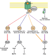
Agonist-induced conformational changes in GPCRs. Agonist-driven conformational changes within the activated GPCR allow exchange of GDP for GTP; Gα-GTP dissociates from Gβγ and stimulates downstream effectors (gray). GPCRs couple to G proteins belonging to four families. The Gα12/13 family activates Rho kinases to stimulate cytoskeletal rearrangements. The Gαq/11 family activates phospholipase C (PLC) to liberate diacylglycerol (DAG) and inositol trisphosphate (IP3) [90]. DAG activates mitogen-activated protein kinase (MAPK) cascades, while IP3 binds to IP3 receptors on the ER to release stored intracellular calcium (Ca2+i). The Gαs family activates adenylate cyclase to increase cAMP which can activate a number of downstream proteins including the enzyme protein kinase A. The Gαi/o family inhibits adenylate cyclase.
Figure 5.
Figure 5.
Amino acid residues required for receptor desensitization and internalization: the dopamine D1 receptor study in Chinese hamster ovary (CHO) cells. The substitution of 256Ser and 257Ser to Ala has been reported to be critical to arrestin-mediated desensitization in HEK293 cells [157]. By contrast, substitution of 359Glu or 360Thr with Ala in CHO cells has been reported to result in desensitization-deficient mutants of the dopamine D1 receptor that are still able to internalize to some extent [153]. Phosphorylation sites in a 12-amino acid stretch of the distal carboxyl tail (428Thr to 439Thr and 446Thr) may be involved in internalization of the receptor in HEK 293 cels [153]. Some studies suggest that the residues in this region of the carboxyl tail may interact with the third loop residues to modulate these effects [158] and that this region may be involved in endocytosis [159].
Figure 6.
Figure 6.
In vitro effects of mutation on desensitization and internalization of the dopamine D1 receptor. Dose-dependent intracellular cAMP accumulation (a and b) and binding curves (c and d) for artificial ligand (SCH 23390) are shown for three constructs: control (wild-type, a and c) and the Thr360Ala mutant (360, b and d). In the desensitization experiments, CHO cells were preincubated with 10 μmol/L dopamine (open circle) or vehicle (closed circle) for 20 min and increasing concentrations of dopamine (10−10 to 10−4 μmol/L) were added to assess cAMP accumulation. Desensitization of the wild-type receptor (a), defined by an increase in Km and decrease in Vmax for agonist-pretreated compared with naïve cells, was abolished (with respect to efficacy and potency) for Thr360Ala (b). Conversely, internalization, defined as a loss of cell surface receptors (measured by decreased maximal binding or Bmax assessed by SCH23390 binding) is unchanged from wild-type (c) after pretreatment with 10 μmol/L dopamine (open circle, compared to vehicle (closed circle), for the Thr360Ala mutation (d) [153].
Figure 7.
Figure 7.
Schematic showing intracellular sites at which GPCR signaling has been described. GPCRs are produced within the biosynthetic pathway and reside in plasma membranes from which they generate G protein signaling. Following receptor internalization via clathrin-coated pits (CCPs) GPCRs can internalize to very early endosomes (VEEs, which are characterized by the presence of leucine zipper 1), or early endosomes, from which additional G protein signaling can be produced. GPCRs including LHR and FFAR2 can signal from VEEs, while receptors including V2R and PTH1R can signal from early endosomes, which may involve β-arrestin (βarr). GPCRs may be trafficked via retromer complexes to the trans-Golgi network. TSHR signals from these sites to generate cAMP and CREB-mediated transcription.

References

    1. Pierce KL, Premont RT, Lefkowitz RJ.. Seven-transmembrane receptors. Nat Rev Mol Cell Biol. 2002;3(9):639–650. doi: 10.1038/nrm908. - DOI - PubMed
    1. Alexander SPH, Christopoulos A, Davenport AP, et al. . The concise guide to pharmacology 2019/20: G protein-coupled receptors. Br J Pharmacol. 2019;176 Suppl 1(Suppl 1):S21–S141. doi: 10.1111/bph.14748. - DOI - PMC - PubMed
    1. Thompson MD. Innovations in phenotyping and diagnostics create opportunities for improved treatment and genetic counseling for rare diseases. Genes (Basel). 2024;15(6):715. doi: 10.3390/genes15060715. - DOI - PMC - PubMed
    1. Hauser AS, Gloriam DE, Bräuner-Osborne H, et al. . Novel approaches leading towards peptide GPCR de-orphanisation. Br J Pharmacol. 2020;177(5):961–968. doi: 10.1111/bph.14950. - DOI - PMC - PubMed
    1. Liu X, Davis D, Segaloff DL.. Disruption of potential sites for N-linked glycosylation does not impair ­hormone binding to the lutropin/choriogonadotropin receptor if Asn-173 is left intact. J Biol Chem. 1993;268(3):1513–1516. - PubMed

MeSH terms

Substances

LinkOut - more resources