Review
. 2025 Jun;22(6):443-455.
doi: 10.1038/s41569-024-01110-1. Epub 2025 Jan 2.

Phenotyping atherosclerotic plaque and perivascular adipose tissue: signalling pathways and clinical biomarkers in atherosclerosis

Affiliations
Review

Phenotyping atherosclerotic plaque and perivascular adipose tissue: signalling pathways and clinical biomarkers in atherosclerosis

Kajetan Grodecki et al. Nat Rev Cardiol. 2025 Jun.

Abstract

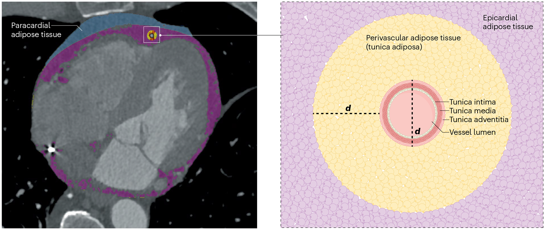
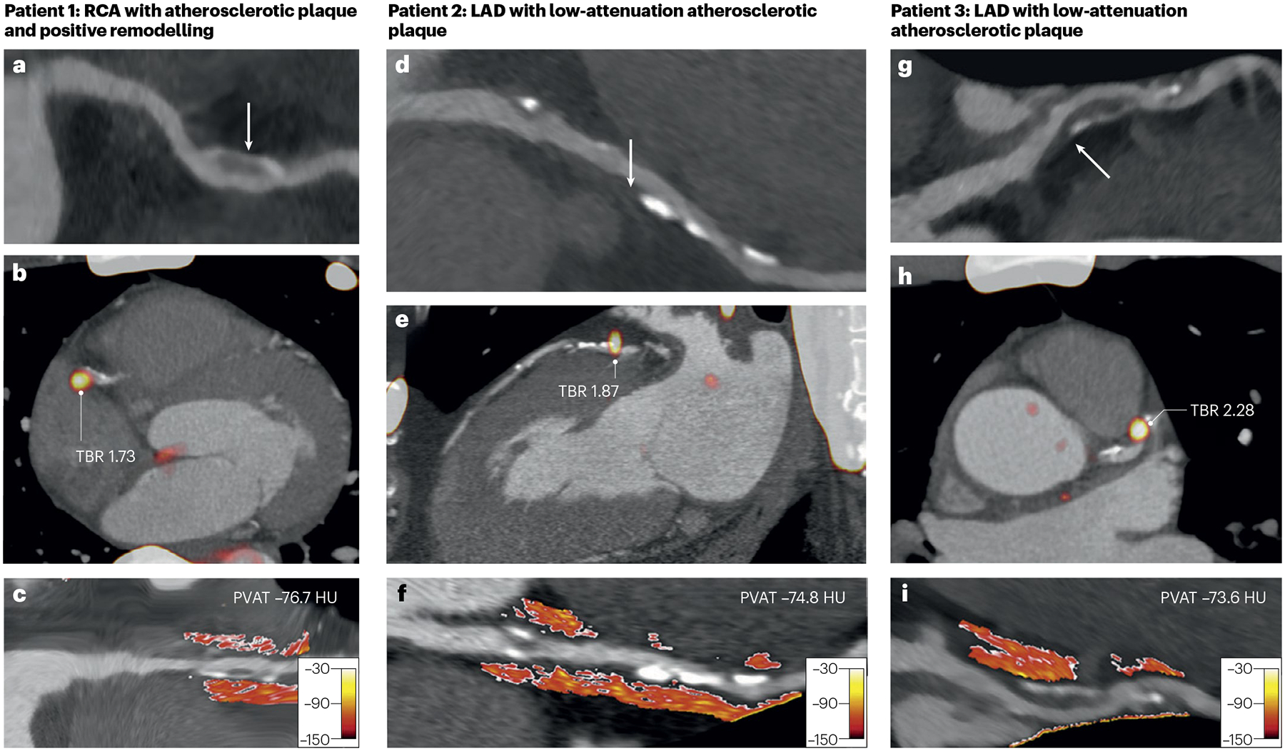
Computed tomography coronary angiography provides a non-invasive evaluation of coronary artery disease that includes phenotyping of atherosclerotic plaques and the surrounding perivascular adipose tissue (PVAT). Image analysis techniques have been developed to quantify atherosclerotic plaque burden and morphology as well as the associated PVAT attenuation, and emerging radiomic approaches can add further contextual information. PVAT attenuation might provide a novel measure of vascular health that could be indicative of the pathogenetic processes implicated in atherosclerosis such as inflammation, fibrosis or increased vascularity. Bidirectional signalling between the coronary artery and adjacent PVAT has been hypothesized to contribute to coronary artery disease progression and provide a potential novel measure of the risk of future cardiovascular events. However, despite the development of more advanced radiomic and artificial intelligence-based algorithms, studies involving large datasets suggest that the measurement of PVAT attenuation contributes only modest additional predictive discrimination to standard cardiovascular risk scores. In this Review, we explore the pathobiology of coronary atherosclerotic plaques and PVAT, describe their phenotyping with computed tomography coronary angiography, and discuss potential future applications in clinical risk prediction and patient management.


Conflict of interest statement

Competing interests: P.J.S., D.B. and D.D. may receive royalties from Cedars-Sinai Medical Center and have equity in APQ Health. The other authors declare no competing interests.

Figures

Fig. 1 |
Fig. 1 |. Anatomy and definitions of thoracic fat.
Paracardial adipose tissue (blue), epicardial adipose tissue (purple) and perivascular adipose tissue (yellow) are thoracic fat depots that can be measured by cardiac computed tomography. Epicardial adipose tissue is located between the myocardium and the visceral layer of the pericardium. Pericardial adipose tissue includes the paracardial and epicardial fat together. Perivascular adipose tissue is defined as being within a radial distance from the outer vessel wall that is equal to vessel diameter (d) and can be considered to be the fourth layer of the artery wall (also referred to as the tunica adiposa).
Fig. 2 |
Fig. 2 |. The PVAT secretome: anti-inflammatory and pro-inflammatory profiles.
a, Under physiological conditions, the secretome of the beige adipocytes in the perivascular adipose tissue (PVAT) includes anti-inflammatory and vasorelaxant compounds. b, Various risk factors, including ageing, obesity, smoking and comorbidities such as diabetes mellitus, result in whitening of adipocytes in the PVAT. This process contributes to the progression of atherosclerosis because the secretome of the PVAT changes to include pro-inflammatory and vasoconstrictive compounds.
Fig. 3 |
Fig. 3 |. Bidirectional interactions between PVAT and the vessel wall in atherosclerosis.
The vessel wall and the perivascular adipose tissue (PVAT) are hypothesized to interact during the formation of atherosclerotic plaques. Adipokines released by the PVAT can affect atherosclerotic plaque formation (outside-to-inside signalling). The same adipocytes might also change in response to the local inflammation of the diseased vessel segment (inside-to-outside signalling). a, Various factors impair endothelial function, primarily through inflammation and oxidative stress. Decreased production of major atheroprotective neurotransmitters, such as nitric oxide and hydrogen sulfide, by endothelial cells is a hallmark of early atherosclerosis. b, Atherosclerotic plaques form through the accumulation of LDL particles in the arterial intima, the infiltration of inflammatory immune cells into the artery wall and the migration of vascular smooth muscle cells to the intima. These cells take up lipoproteins and transform into foam cells. In response to changes in the vessel wall, PVAT starts secreting pro-inflammatory cytokines that promote further infiltration of foam cell precursors into the intimal layer. c, Atherosclerosis progression leads to the formation of a vulnerable plaque with a large thrombogenic necrotic core covered by a thin layer of fibrous tissue. Excess accumulation of necrotic cells and cholesterol crystals contributes to growth of the necrotic core. Moreover, a pro-inflammatory environment downregulates the expression of genes responsible for collagen synthesis by vascular smooth muscle cells, contributing to plaque vulnerability and decreasing the thickness of the fibrous cap.
Fig. 4 |
Fig. 4 |. Associations between 18F-NaF uptake and PVAT in coronary artery disease.
Increased atherosclerotic plaque vulnerability measured by 18F-NaF uptake (a marker of active calcification) on positron emission tomography and by attenuation of perivascular adipose tissue (PVAT) on computed tomography coronary angiography. Patient 1 is a man aged 53 years with right coronary artery (RCA) atherosclerotic plaque with positive remodelling (arrow) (part a), focal 18F-NaF uptake with an elevated target-to-background ratio (TBR) of 1.73 (part b), and increased PVAT attenuation (mean PVAT density −76.7 Hounsfield units (HU)) (part c). Patient 2 is a man aged 66 years with left anterior descending coronary artery (LAD) atherosclerotic plaque with low attenuation (arrow) (part d), focal 18F-NaF uptake with an elevated TBR of 1.87 (part e) and increased PVAT attenuation (mean PVAT density −74.8 HU) (part f). Patient 3 is a man aged 54 years with LAD atherosclerotic plaque with low attenuation (arrow) (part g), focal 18F-NaF uptake with an elevated TBR of 2.28 (part h) and increased PVAT attenuation (mean PVAT density −73.6 HU) (part i). Adapted with permission from ref. , Elsevier.
Fig. 5 |
Fig. 5 |. Atherosclerotic plaque regression and stabilization.
Treatment of a patient with 20 mg of rosuvastatin led to a 44% decrease in plasma LDL-cholesterol levels between baseline (part a) and the 3-year follow-up (part b). Computed tomography coronary angiography shows that the amount of non-calcified and low-attenuation atherosclerotic plaque decreased, whereas the amount of calcified atherosclerotic plaque increased, thereby increasing plaque stability. The mean attenuation of the perivascular adipose tissue decreased by 2 Hounsfield units.

References

    1. Vaduganathan M, Mensah GA, Turco JV, Fuster V & Roth GA The global burden of cardiovascular diseases and risk. J. Am. Coll. Cardiol 80, 2361–2371 (2022). - PubMed
    1. Ibanez B et al. Progression of early subclinical atherosclerosis (PESA) study. J. Am. Coll. Cardiol 78, 156–179 (2021). - PubMed
    1. Libby P et al. Atherosclerosis. Nat. Rev. Dis. Prim 5, 56 (2019). - PubMed
    1. Arbab-Zadeh A, Nakano M, Virmani R & Fuster V Acute coronary events. Circulation 125, 1147–1156 (2012). - PMC - PubMed
    1. Falk E, Shah PK & Fuster V Coronary plaque disruption. Circulation 92, 657–671 (1995). - PubMed

MeSH terms

LinkOut - more resources